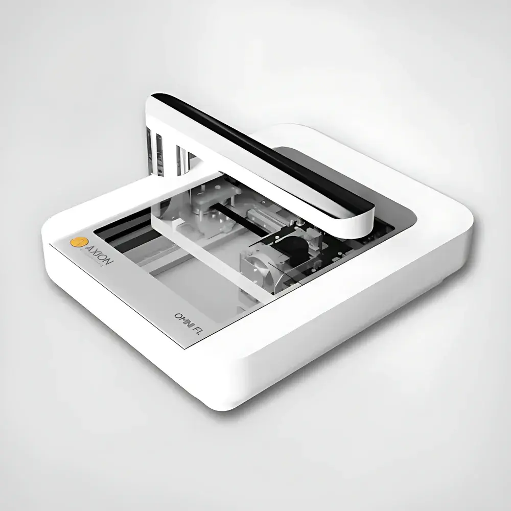
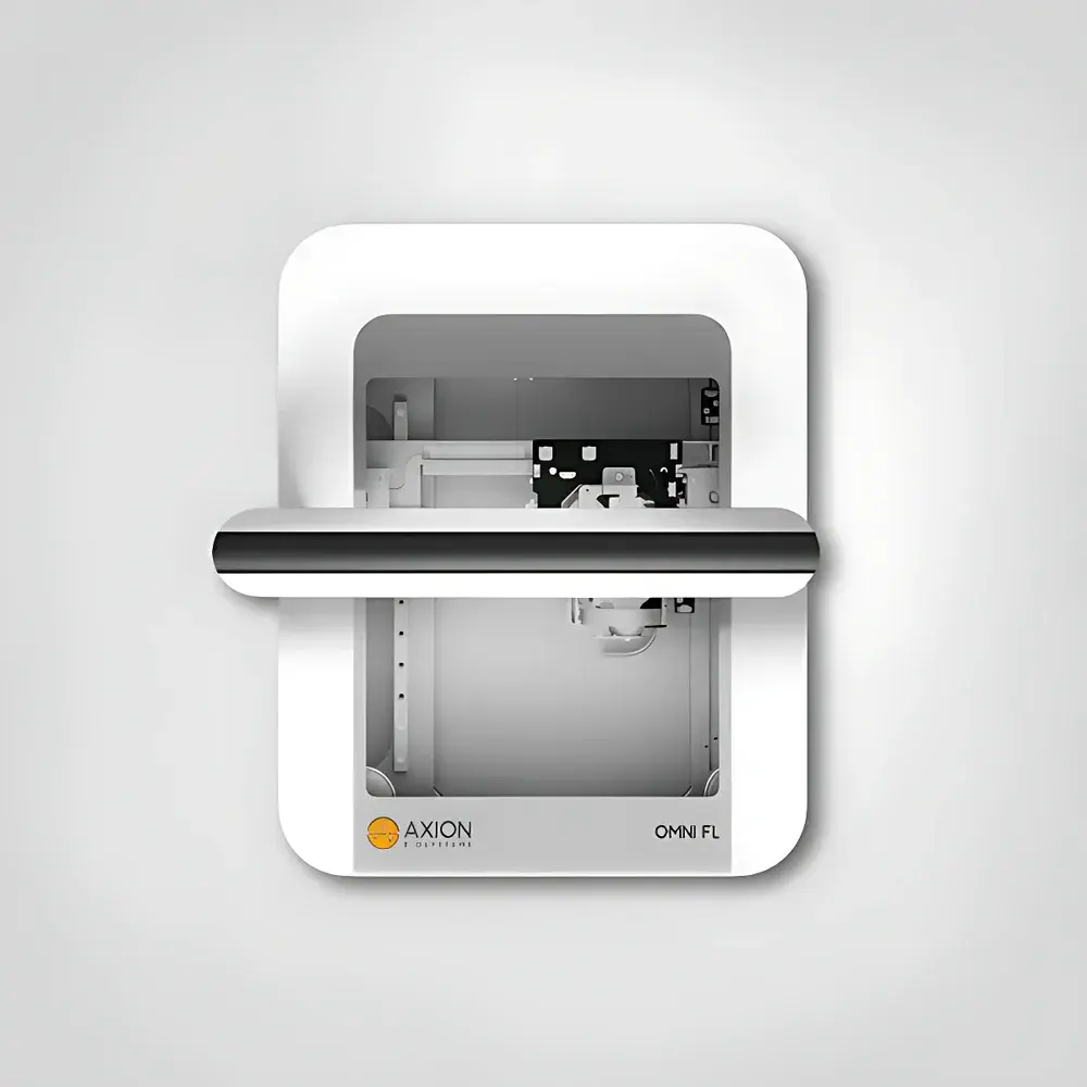
Axion BioSystems Omni In-Cell Incubator-Based Live-Cell Imaging and Analysis System
| Brand | Axion BioSystems |
|---|---|
| Origin | Netherlands |
| Manufacturer Type | Original Equipment Manufacturer (OEM) |
| Origin Category | Imported |
| Model | Omni / Lux3 |
| Pricing | Available Upon Request |
Overview
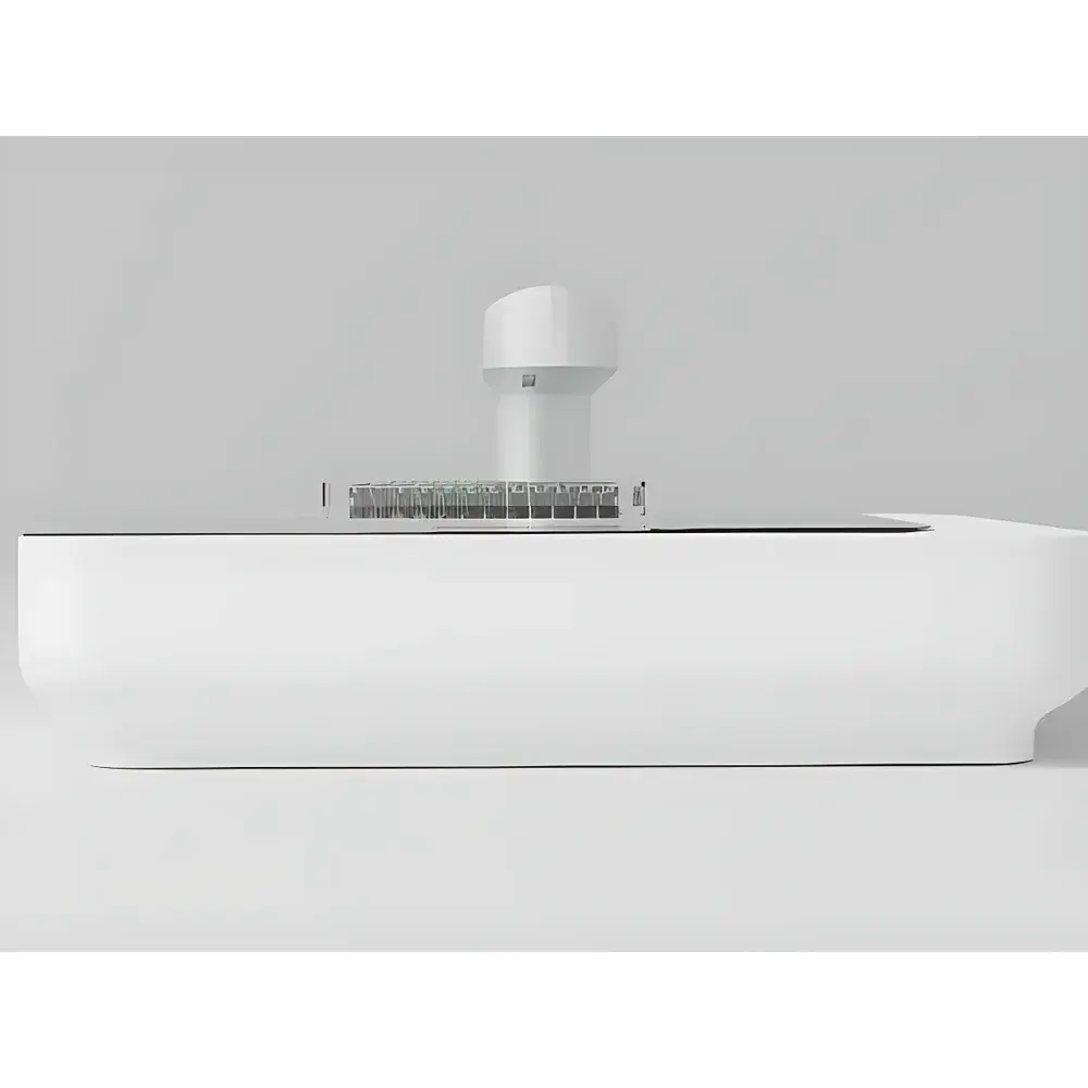
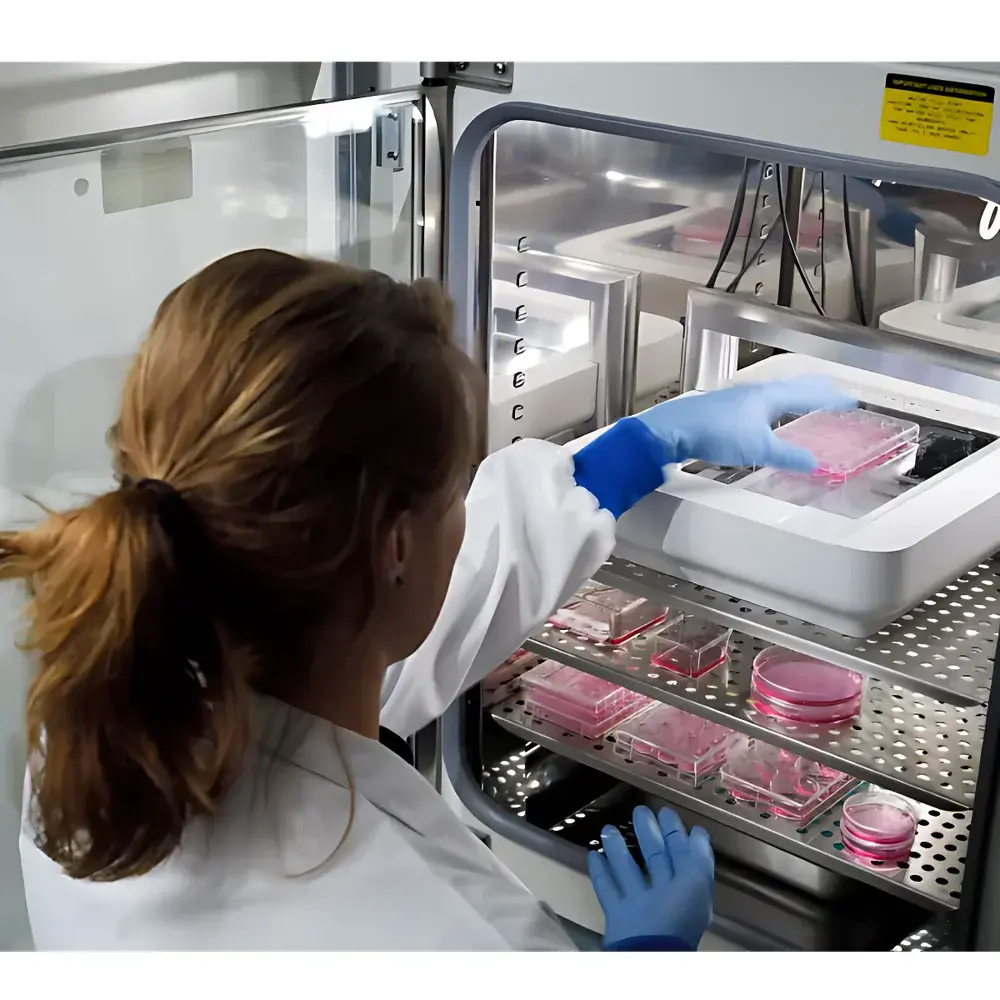
The Axion BioSystems Omni is an incubator-integrated, label-free live-cell imaging platform engineered for longitudinal, non-invasive monitoring of cellular dynamics under physiologically relevant conditions. Unlike conventional benchtop microscopes requiring sample removal from controlled environments, the Omni operates entirely within standard CO2 incubators (5–40 °C, 20–95% RH), preserving native cell behavior by eliminating thermal, humidity, and pH perturbations associated with external handling. Its core architecture combines high-resolution brightfield imaging with optional fluorescence excitation (470 nm and 590 nm LED sources) to support both label-free morphological assessment and targeted fluorescent reporter assays. The system employs a motorized objective lens assembly that traverses beneath the sample stage—enabling precise, coordinate-driven scanning across multi-well plates without mechanical displacement of vessels. This design ensures measurement integrity across time-series experiments, particularly critical for applications demanding high temporal fidelity, such as proliferation kinetics, cytotoxicity profiling, and immune cell–target engagement assays.
Key Features
- In-incubator operation: Fully validated for continuous deployment inside standard humidified CO2 incubators; all electronics and optics rated for sustained operation at 5–40 °C and up to 95% relative humidity.
- Dual-mode optical detection: Simultaneous brightfield and optional fluorescence (green: ~470 nm excitation; red: ~590 nm excitation) channels, enabling comparative analysis of unlabeled morphology and labeled functional responses.
- Full-plate brightfield tiling: Automated acquisition of up to ~7,850 individual high-resolution frames per scan; software-based stitching yields a seamless composite image (86 mm × 124 mm), covering the entire usable field of view.
- Flexible vessel compatibility: Supports any transparent culture vessel ≤55 mm in height—including 6- to 384-well plates, Petri dishes, and T25–T225 flasks—without adapter requirements or recalibration.
- Zero-intervention imaging workflow: No plate movement, no manual focusing, no daily alignment; factory-calibrated optics and embedded autofocus routines ensure consistent image quality across weeks-long experiments.
- Cloud-connected data pipeline: Raw image stacks uploaded automatically to CytoSMART Cloud, supporting secure remote access, audit-trail-enabled analysis, and integration with local or third-party computational tools.
Sample Compatibility & Compliance
The Omni accommodates standard ANSI/SLAS-compliant microplates (SBS format), glass-bottom dishes, and custom microfluidic devices, provided optical clarity and dimensional constraints (≤55 mm height, ≤86 mm × 124 mm footprint) are met. All hardware components comply with IEC 61000-6-3 (EMC emission standards) and IEC 61010-1 (safety requirements for laboratory equipment). Data management workflows align with GLP/GMP expectations through timestamped metadata embedding, user-access logging, and exportable audit trails—facilitating regulatory submissions under FDA 21 CFR Part 11 when paired with appropriate institutional IT validation protocols.
Software & Data Management
CytoSMART Software provides intuitive experiment setup, real-time monitoring dashboards, and modular algorithm-based analysis. Pre-validated modules include confluence quantification (brightfield and fluorescence), scratch/wound-healing migration tracking, colony formation enumeration, and fluorescent object counting. Each module generates traceable output files (.csv, .tif, .json) containing raw pixel values, segmentation masks, and statistical summaries. Users retain full ownership of raw data and may export unprocessed TIFF stacks for offline analysis in ImageJ/Fiji, MATLAB, or Python-based pipelines (e.g., scikit-image, CellProfiler). All cloud interactions use TLS 1.2+ encryption; on-premise deployment options are available for institutions requiring air-gapped infrastructure.
Applications
- Tumor spheroid modeling: Long-term monitoring of 3D aggregate formation, compaction, and drug-induced disintegration—supporting oncology compound screening aligned with ISO 10993-5 cytotoxicity guidelines.
- Proliferation & viability kinetics: Label-free confluence metrics correlated with metabolic activity (e.g., ATP assays) to deconvolute growth arrest vs. cytostatic effects.
- Immuno-oncology assays: Real-time quantification of CAR-T or NK cell–mediated tumor lysis, including synapse formation dynamics and target cell membrane integrity loss.
- Mechanistic toxicology: Time-resolved assessment of mitochondrial depolarization (using TMRE), calcium flux (Fluo-4), or nuclear condensation (Hoechst) in response to small molecules or biologics.
- Transfection efficiency validation: Co-localized brightfield morphology and fluorescent protein expression to distinguish transfection success from cytotoxic side effects.
- Migration and invasion assays: Automated wound closure rate calculation with spatial normalization across multi-well replicates—reducing inter-operator variability inherent in manual scratch assays.
FAQ
How does the Omni acquire images without moving the culture plate?
The system uses a precision-guided, motorized objective lens positioned beneath the sample stage. During brightfield scanning, the lens moves in a grid pattern while the plate remains stationary—capturing overlapping sub-images that are computationally stitched into a single high-fidelity mosaic.
What fluorescence wavelengths are supported?
The optional FL Module includes two narrow-band LED sources: 470 nm (optimized for GFP, FITC, Calcein-AM) and 590 nm (optimized for RFP, mCherry, Propidium Iodide). Excitation/emission filtering is integrated into the optical path.
Can I perform kinetic assays over several days?
Yes. The Omni supports scheduled imaging intervals from 1 minute to 24 hours, with battery-backed real-time clock synchronization and redundant storage buffering to prevent data loss during brief incubator door openings.
Is calibration required before each experiment?
No. The system undergoes factory calibration for focus, illumination uniformity, and stage positioning. Auto-focus routines run prior to each scan to compensate for minor thermal drift.
Which file formats are generated?
Raw data exports as multi-page TIFF stacks; processed outputs include CSV tables (quantitative metrics), JSON metadata logs, and annotated PNG overlays—all compliant with FAIR data principles.


