Axion BioSystems Omni FL Live-Cell Imaging System for 3D Tumor Spheroid Monitoring
| Brand | Axion BioSystems |
|---|---|
| Origin | Netherlands |
| Manufacturer | Axion BioSystems |
| Product Type | Imported |
| Model | Omni FL |
| Temperature Control Range | 5–40 °C |
| Humidity Control Range | 20–95% RH |
| Full-Plate Imaging Speed | 10 min per plate |
Overview
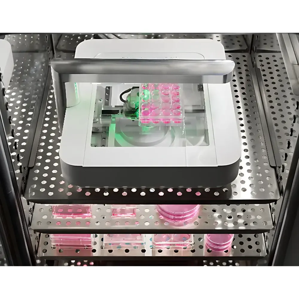
The Axion BioSystems Omni FL is a compact, integrated live-cell imaging workstation engineered for long-term, label-free and fluorescence-based monitoring of 3D tumor spheroids and organoids inside standard CO₂ incubators. Leveraging wide-field LED illumination and high-resolution CMOS imaging, the system operates on the principle of time-lapse brightfield and epifluorescence microscopy—optimized for non-invasive, longitudinal observation under physiologically relevant culture conditions. Unlike conventional benchtop microscopes, the Omni FL mounts directly within the incubator, eliminating thermal and pH perturbations associated with sample removal. Its optical architecture supports quantitative morphometric analysis of spheroid formation kinetics, compaction dynamics, size distribution, and viability-associated structural changes—enabling rigorous in vitro modeling of solid tumor biology, stromal interactions, and therapeutic response heterogeneity.
Key Features
- Incubator-integrated design: Fully functional at 5–40 °C and 20–95% RH; no external hardware or heat dissipation compromises
- Dual-mode imaging: Simultaneous brightfield and fluorescence (GFP, RFP, and other standard fluorophores) acquisition with programmable exposure control
- Full-plate scanning capability: Automated mosaic imaging across 6–384-well plates, generating stitched panoramas up to 86 mm × 124 mm (effective field)
- Rapid acquisition protocol: Complete brightfield scan of a 96-well plate in ≤10 minutes; configurable z-stack and multi-position fluorescence capture per well
- Modular software architecture: Cloud-hosted image storage and processing via CytoSMART Cloud; local data export in TIFF/OME-TIFF format for compliance with FAIR data principles
- Robust mechanical stability: Precision motorized stage and fixed LED source minimize vibration-induced artifacts during extended time-lapse experiments
Sample Compatibility & Compliance
The Omni FL accommodates all optically transparent, flat-bottomed culture vessels with height <55 mm—including 6-, 24-, 48-, 96-, and 384-well plates, Petri dishes, and T25–T225 flasks. It is validated for use with primary tumor-derived spheroids, patient-derived organoids (PDOs), co-cultures (e.g., tumor-immune or tumor-stroma), and genetically engineered models (e.g., GFP-labeled HeLa/C6 co-cultures). The system conforms to ISO 13485 design controls for medical device-related research instrumentation and supports audit-ready workflows compliant with GLP and GMP-aligned documentation standards. Image metadata includes timestamp, environmental log (temperature/humidity), objective settings, and user-defined experimental annotations—ensuring traceability per FDA 21 CFR Part 11 requirements when paired with appropriate access controls.
Software & Data Management
All images are automatically uploaded to the encrypted CytoSMART Cloud platform, where users access them via browser-based interface. Pre-installed analysis modules include: (1) Brightfield/fluorescence confluence quantification, (2) Spheroid segmentation and size distribution profiling (area, perimeter, circularity, solidity), (3) Scratch/wound healing migration tracking, (4) Clonogenic assay counting and growth curve derivation, and (5) Fluorescent object counting (e.g., CAR-T cell infiltration events). Raw OME-TIFF stacks can be downloaded for advanced analysis in Fiji/ImageJ, MATLAB, or Python-based pipelines (e.g., CellProfiler, DeepCell). Audit trails record user actions, parameter modifications, and analysis versioning—supporting reproducibility and regulatory submission readiness.
Applications
- Tumor spheroid development & drug response: Quantify growth inhibition, necrosis onset, and morphological collapse following chemotherapy, targeted therapy, or immune effector exposure
- Immunooncology assays: Monitor real-time CAR-T or NK cell–mediated spheroid disintegration, including spatial metrics of immune synapse formation and cytolytic penetration depth
- Stromal-tumor interaction modeling: Track fibroblast-driven spheroid compaction or endothelial sprouting in co-culture formats
- Toxicity screening: Perform kinetic assessment of compound-induced cytotoxicity without endpoint dyes—preserving samples for downstream omics
- Clonogenic survival assays: Automate colony identification and expansion kinetics across entire plates under continuous incubation
- Transfection/transduction efficiency: Correlate fluorescent reporter expression onset with morphological differentiation in stem-like spheroids
FAQ
How does the Omni FL acquire images inside an incubator?
The system uses a fixed top-mounted LED light source and a motorized bottom-stage camera that moves beneath the plate. In brightfield mode, it captures overlapping tiles across the full plate surface; software stitches them into a single high-resolution panorama. In fluorescence mode, users define specific well positions and exposure parameters per channel.
What image analysis algorithms are available?
Pre-validated modules include confluence analysis, spheroid segmentation, scratch assay migration tracking, clonogenic counting, and fluorescent object detection. Custom analysis is supported via raw TIFF/OME-TIFF export.
Can the Omni FL be used outside a CO₂ incubator?
Yes—it operates across 5–40 °C and 20–95% RH, making it suitable for hypoxia chambers, room-temperature studies, or laminar flow hoods with environmental control.
Which plate formats are supported?
All standard flat-bottom, optically clear plates from 6- to 384-well, plus Petri dishes and T-flasks up to 55 mm in height. Effective imaging area is limited to 86 mm × 124 mm per scan.
Is data export compatible with LIMS or ELN systems?
Yes—structured metadata (JSON/XML) and OME-TIFF files support integration with laboratory information management systems (LIMS) and electronic lab notebooks (ELN) via REST API or SFTP protocols.


