Bio-Rad ChemiDoc High-Sensitivity Chemiluminescence Imaging System
| Brand | Bio-Rad |
|---|---|
| Origin | Singapore |
| Model | ChemiDoc |
| Instrument Type | Chemiluminescence Gel Imaging System |
| CCD Resolution | 6.1 MP |
| Bit Depth | 16-bit |
| Dynamic Range | 0–4.8 OD |
| CCD Sensor Size | 1-inch |
| Detection Sensitivity | fg-level protein |
| Signal-to-Noise Ratio | >56 dB |
| Lens | Fixed-focus |
Overview
The Bio-Rad ChemiDoc High-Sensitivity Chemiluminescence Imaging System is a dedicated, cooled CCD-based imaging platform engineered for quantitative detection of chemiluminescent signals from Western blots, nucleic acid gels, and other low-light biological assays. Unlike traditional film-based autoradiography or X-ray film development—which require darkroom infrastructure, chemical developers, fixers, and manual exposure optimization—the ChemiDoc system captures high-fidelity luminescent emissions directly via a thermoelectrically cooled 6.1-megapixel CCD sensor. Its optical architecture leverages a fixed-focus lens calibrated across all sample heights and magnification settings, ensuring consistent spatial resolution without manual refocusing. The system operates on the principle of photon integration over user-defined exposure durations, with real-time preview enabling rapid protocol iteration. Designed for routine laboratory environments, it delivers reproducible quantitation across wide dynamic ranges (0–4.8 optical density units), supporting rigorous comparative analysis in molecular biology, immunoblotting, and functional proteomics workflows.
Key Features
- Thermoelectrically cooled 1-inch CCD sensor with 16-bit digitization for high-resolution signal capture and minimal thermal noise
- fg-level detection sensitivity for low-abundance proteins—validated against standard horseradish peroxidase (HRP)-conjugated secondary antibodies
- Auto-detection of application-specific trays (e.g., mini-gel, large-format blot, or multi-well plate) to load preconfigured acquisition parameters
- Two dedicated chemiluminescence acquisition modes: “Quick Capture” for rapid screening and “Auto Optimize Exposure” for iterative signal maximization within dynamic range limits
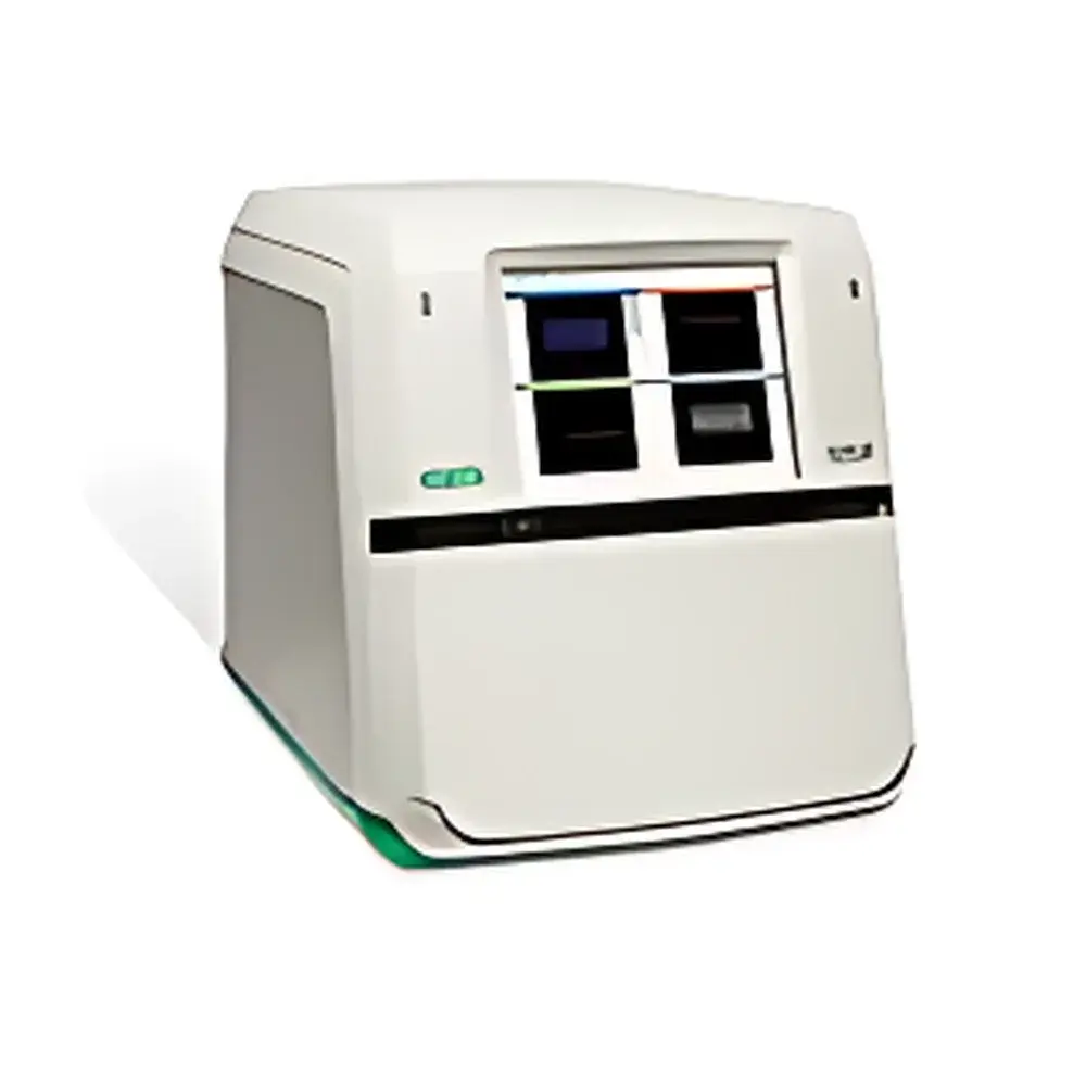
- 12.1-inch multi-touch LCD display with intuitive gesture-based interface for real-time image preview, region-of-interest (ROI) definition, and on-device annotation
- Pre-calibrated optical path ensures focus consistency across variable sample heights—from thin transfer membranes to thick agarose gels—eliminating manual focus drift during throughput runs
- Modular design supports field-upgradeability to the ChemiDoc MP platform, extending functionality to fluorescence, UV transillumination, and white-light documentation
Sample Compatibility & Compliance
The ChemiDoc system accommodates standard electrophoretic formats including mini- and midi-gels (up to 15 × 15 cm), nitrocellulose and PVDF membranes, multi-well plates (6–96-well), and stained or unstained nucleic acid gels. Its hardware and software stack comply with core regulatory expectations for data integrity in GLP- and GMP-aligned laboratories: audit-trail-enabled acquisition logs, user-access controls, and timestamped metadata embedding (exposure time, gain, binning, lens position, tray ID). While not inherently 21 CFR Part 11 compliant out-of-the-box, the system integrates seamlessly with Bio-Rad’s Image Lab Touch Software—available in validated configurations that support electronic signatures, role-based permissions, and secure data export in TIFF, PNG, and vendor-neutral XML-based formats compliant with ISO/IEC 17025 documentation standards.
Software & Data Management
Controlled by Bio-Rad’s Image Lab Touch Software, the ChemiDoc platform provides integrated tools for background subtraction, lane/strip detection, band intensity quantification, molecular weight estimation, and normalization against loading controls. All processing steps are recorded in a non-erasable acquisition log, preserving traceability from raw image to final graph. Data export supports batch processing via scripting (Python API available), direct import into statistical packages (e.g., GraphPad Prism, R), and LIMS-compatible CSV/TXT outputs. Image calibration is maintained through factory-installed reference standards, enabling inter-instrument comparability when deployed across multi-site research or QC labs. Raw image files retain full 16-bit depth and embedded EXIF-like metadata—including sensor temperature, exposure duration, and analog gain—ensuring full analytical reproducibility.
Applications
- Quantitative Western blot analysis using HRP- or AP-based chemiluminescent substrates (e.g., ECL, CDP-Star)
- Detection of low-copy-number phosphoproteins and post-translational modifications under stringent washing conditions
- High-throughput screening of antibody specificity and titration curves
- Validation of CRISPR/Cas9 editing efficiency via cleavage product quantification
- Comparative expression profiling across tissue lysates, cell line panels, or time-course experiments
- Documentation and analysis of multiplexed immunoassays on membrane arrays
FAQ
Does the ChemiDoc system require darkroom conditions for chemiluminescent imaging?
No. The system’s light-tight imaging chamber and cooled CCD eliminate ambient light interference, enabling operation in standard laboratory lighting.
Can I perform fluorescence imaging on the base ChemiDoc model?
No—fluorescence capability requires upgrade to the ChemiDoc MP configuration, which includes LED excitation sources and emission filters.
Is the 16-bit image data preserved throughout processing?
Yes. All quantitative operations (background correction, normalization, densitometry) operate on the full 16-bit pixel array; exported results retain original bit-depth fidelity unless explicitly down-sampled.
How is calibration maintained across instruments or over time?
Each unit ships with NIST-traceable optical density step tablets and internal reference LEDs. Routine verification protocols are built into Image Lab Touch and generate PDF reports compliant with ISO/IEC 17025 clause 6.4.10.
What file formats are supported for data exchange with third-party analysis tools?
TIFF (uncompressed, 16-bit), PNG (lossless), and XML-based .rdcf files containing raw pixel data plus structured metadata—fully compatible with open-source tools such as Fiji/ImageJ and commercial platforms like PerkinElmer’s Spotfire.


