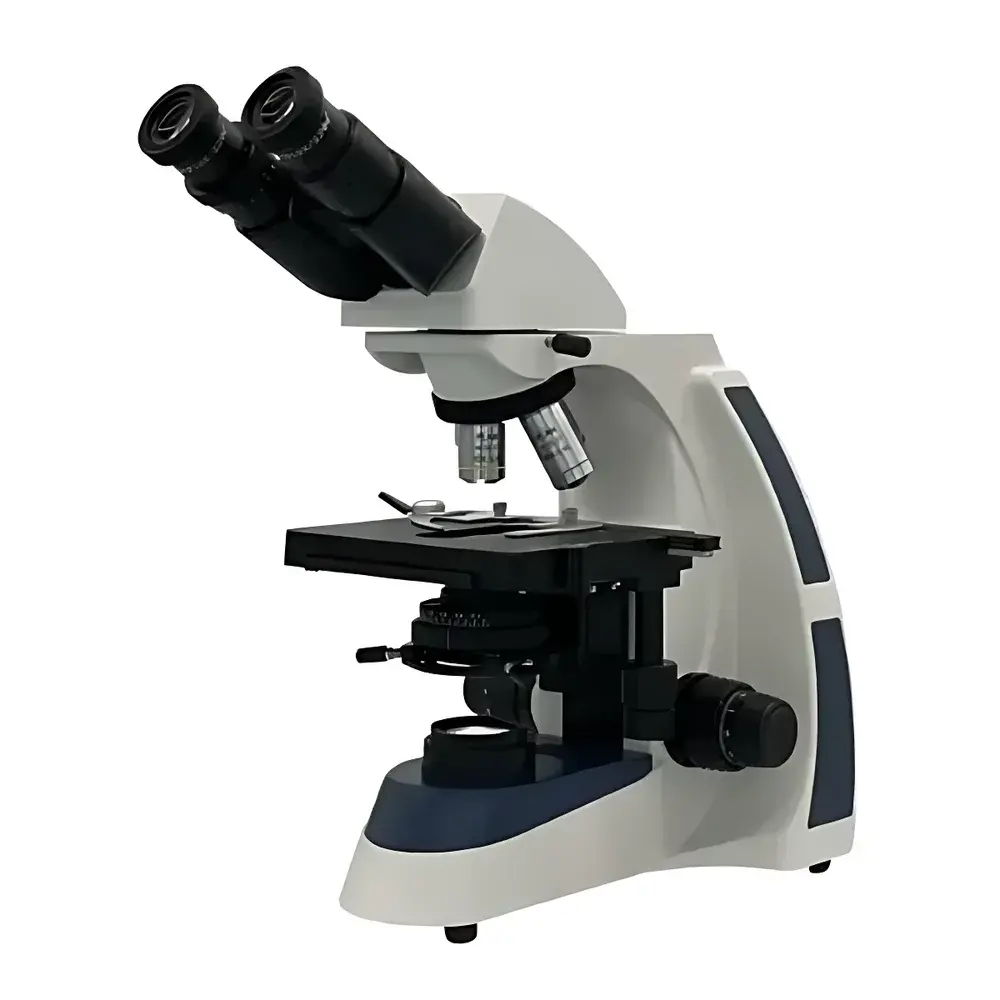

BM Microscopes XSP-BM-17 Upright Biological Microscope
| Brand | BM Microscopes |
|---|---|
| Origin | Shanghai, China |
| Model | XSP-BM-17 |
| Microscope Type | Upright |
| Optical Design | Infinity-Corrected (UIS) |
| Eyepiece Configuration | Binocular |
| Standard Eyepieces | Widefield 10×/Φ22 mm |
| Optional Eyepieces | 16×/Φ15 mm |
| Objective Set | Infinity Plan Achromatic 4× (NA 0.10, WD 20.00 mm), 10× (NA 0.25, WD 13.50 mm), 40× (NA 0.65, WD 0.68 mm), 100× Oil (NA 1.25, WD 0.20 mm) |
| Total Magnification Range | 40×–1000× |
| Mechanical Stage | 140 × 160 mm, Travel Range 76 × 45 mm |
| Focusing System | Coaxial coarse/fine focus with 30 mm travel |
| Fine Focus Graduation | 2 µm |
| Condenser | Abbe condenser, NA 1.25, adjustable iris diaphragm |
| Filters | Blue and green interference filters |
| Illumination | Adjustable-intensity LED light source |
| Tube Angle | 30° inclined, 360° rotatable hinge joint |
| Compliance | Designed for ISO 10993-compatible lab environments |
Overview
The BM Microscopes XSP-BM-17 is an upright biological microscope engineered for precision life science applications requiring high optical fidelity, ergonomic usability, and modular expandability. Built upon an infinity-corrected UIS (Universal Infinity System) optical architecture, the XSP-BM-17 delivers consistent image quality across its full magnification range (40× to 1000×) while supporting future integration of advanced modules—including fluorescence illumination units, phase contrast sliders, and vertical incident light paths. Its rigid mechanical design incorporates a 30° inclined, fully rotatable binocular head optimized for extended observation sessions, and a thermally stable LED illumination system offering flicker-free, color-accurate white light with continuous brightness control—critical for photometric consistency in comparative histological or cytological analysis.
Key Features
- Infinity-corrected plan achromatic objective suite (4×, 10×, 40×, 100× oil) delivering high flatness and chromatic correction across the entire field of view (Φ22 mm)
- Binocular viewing head with interpupillary adjustment (55–75 mm) and diopter compensation on both eyepieces for individual user calibration
- High-precision coaxial focusing mechanism with 30 mm coarse travel and 2 µm fine-focus graduation, enabling reproducible Z-axis positioning for serial section imaging or z-stack acquisition
- Large mechanical stage (140 × 160 mm) with vernier-calibrated X-Y translation (76 × 45 mm range), compatible with standardized specimen slides (76 × 26 mm) and petri dish holders
- Abbe condenser (NA 1.25) with centerable iris diaphragm and swing-out top lens—optimized for Köhler illumination alignment and critical aperture control in brightfield and darkfield modalities
- Dual-band interference filters (blue and green) integrated into the filter holder for spectral selection in unstained or dye-assisted visualization
- Low-heat, long-life LED illumination (≥25,000 h rated lifetime) with linear dimming circuitry—eliminating thermal drift during time-lapse observation and reducing specimen phototoxicity
Sample Compatibility & Compliance
The XSP-BM-17 accommodates standard glass microscope slides (1 × 3 inches), coverslips (No. 1.5, 0.17 mm thickness), Petri dishes (up to 100 mm diameter), and tissue culture flasks via optional stage adapters. Its optical path is calibrated for use with immersion oil (n = 1.515) at 100× magnification, meeting ISO 8578 requirements for oil-immersion resolution validation. The instrument complies with IEC 61010-1:2010 safety standards for laboratory electrical equipment and supports traceable calibration protocols required under GLP (Good Laboratory Practice) and ISO/IEC 17025 frameworks. While not FDA 510(k)-cleared as a diagnostic device, its optical performance aligns with ASTM E2872-22 guidelines for routine biological microscopy in research and academic settings.
Software & Data Management
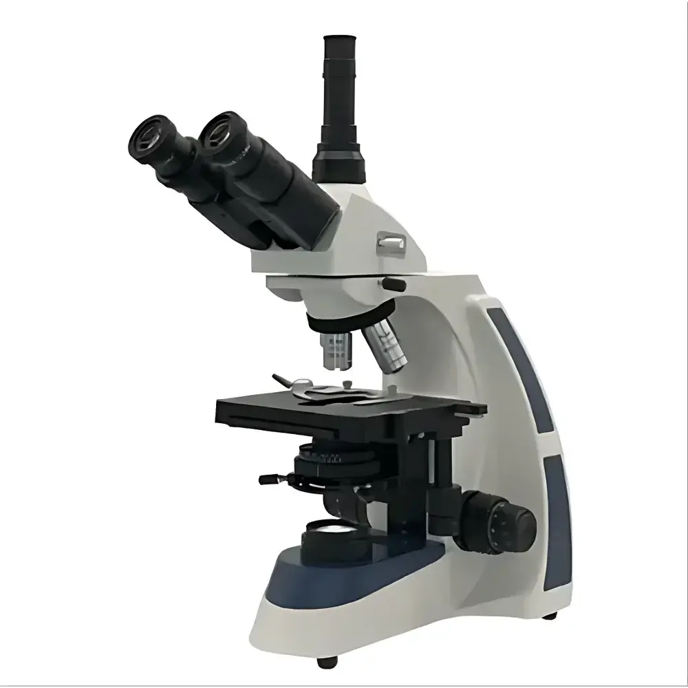
The XSP-BM-17 is designed for seamless integration with third-party digital imaging systems. Its trinocular port (optional accessory) accepts C-mount adapters (0.5× or 1× reduction) for coupling to CMOS/CCD cameras compliant with USB3/UVC or GigE Vision standards. Image acquisition, annotation, measurement (length, area, particle count), and multi-channel overlay are supported via open-API platforms including NIS-Elements (Nikon), ZEN (Zeiss), and open-source tools such as Fiji/ImageJ. Audit trails, user access logs, and metadata embedding (EXIF + custom tags) can be implemented through institutional LIMS or ELN systems—ensuring compliance with 21 CFR Part 11 when paired with validated software configurations.
Applications
This microscope serves as a foundational platform in academic teaching laboratories for introductory histology and microbiology instruction, where robustness and ease of alignment reduce operational overhead. In research contexts, it supports routine tasks including bacterial morphology assessment (Gram staining), mitotic index quantification in cultured cell lines, hematoxylin-eosin (H&E) tissue section evaluation, and basic immunofluorescence screening when equipped with appropriate excitation/emission filters. Its mechanical stability and repeatable focusing make it suitable for longitudinal studies involving live-cell observation (e.g., wound-healing assays in low-magnification brightfield), provided environmental controls (temperature/humidity) are externally maintained.
FAQ
Is the XSP-BM-17 compatible with fluorescence imaging?
Yes—when fitted with a vertical illuminator module (sold separately) and appropriate excitation/emission filter sets, the UIS optical path maintains transmission efficiency above 85% across 365–650 nm.
What is the maximum usable magnification with the standard 10× eyepieces?
The theoretical limit is 1000× (100× oil objective × 10× eyepiece); practical resolution is constrained by the Rayleigh criterion and specimen preparation quality—notably, diffraction-limited resolution is ~0.25 µm at 550 nm wavelength.
Can the LED illumination intensity be controlled externally?
Yes—the rear-panel potentiometer allows analog dimming; for automated control, the DC power input (12 V) supports PWM-driven external controllers via optional interface cable.
Does the microscope include a camera port?
The base configuration includes a monocular viewing tube; a trinocular head with 20:80 beam splitter is available as a factory-installed option or field-upgrade kit.
Are replacement objectives and eyepieces interchangeable with other BM UIS-series microscopes?
Yes—all BM UIS-compatible optics (designated “∞/180”) share standardized thread specifications (RMS 20.32 mm) and parfocal distance (45 mm), ensuring cross-platform compatibility within the BM UIS ecosystem.
Related Products


