
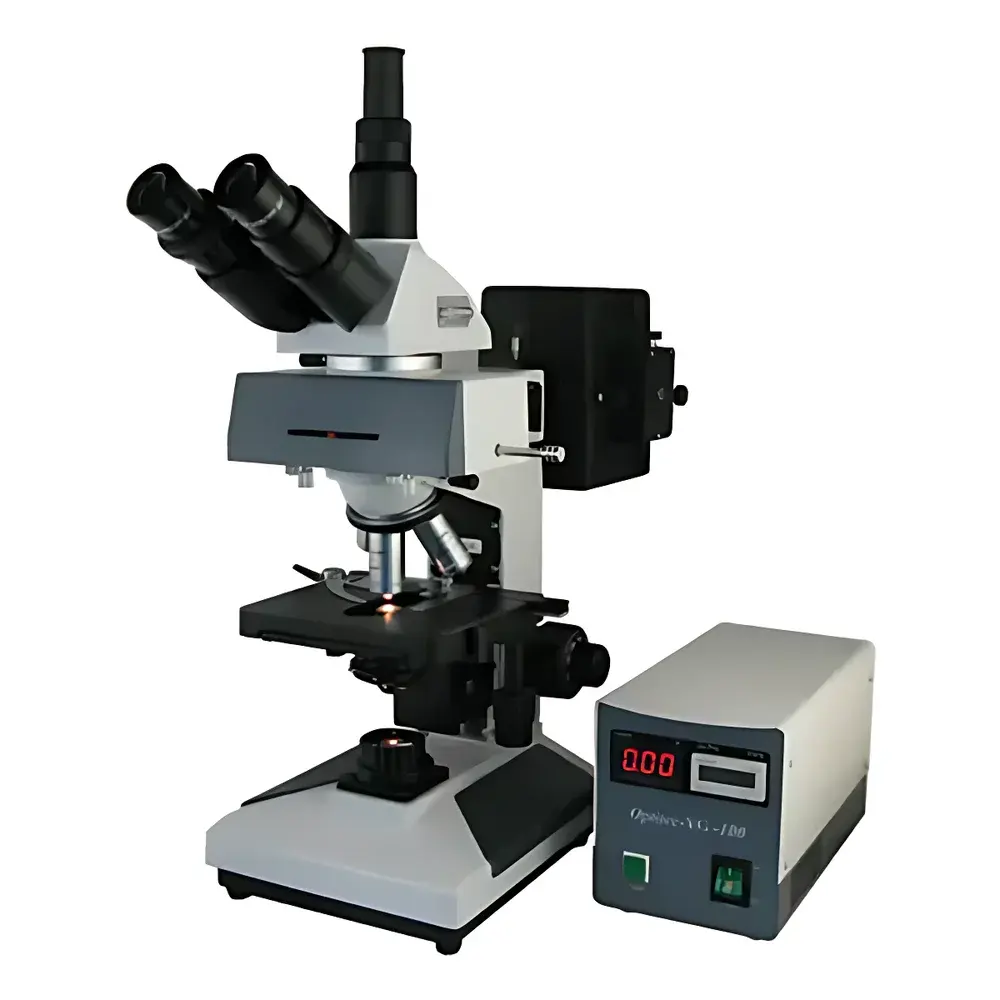

BM XSP-BM-13C Upright Fluorescence Microscope
| Brand | BM |
|---|---|
| Origin | Shanghai, China |
| Model | XSP-BM-13C |
| Instrument Type | Upright Fluorescence Microscope |
| Excitation Source | Mercury Arc Lamp (100 W) with B/G Filter Sets |
| Transmitted Illumination | 6 V / 20 W Halogen Lamp |
| Eyepieces | 10× / Ø20 mm (standard), optional 16× / Ø15 mm |
| Objective System | Infinity-Corrected Fluorescence Objectives (4×, 10×, 40×, 100× oil, NA up to 1.25) |
| Mechanical Stage | 180 × 150 mm platform, 75 × 50 mm travel range |
| Condenser | Adjustable Abbe condenser, NA 1.25 with iris diaphragm |
| Observation Head | 30° inclined binocular head with 360° rotation capability |
| Focusing Mechanism | Coaxial coarse/fine focus, 30 mm vertical travel, fine focus graduation 0.002 mm, tension adjustment and limit stop |
| Fluorescence Filters | Blue (B), Green (G), Yellow (Y) excitation/emission sets |
| Interpupillary Distance | 55–75 mm |
| Tube Factor | 1× (standard infinity system) |
Overview
The BM XSP-BM-13C is an upright fluorescence microscope engineered for routine epifluorescence imaging in academic, clinical, and industrial life science laboratories. It operates on the principle of epifluorescence illumination—where excitation light is directed through the objective lens onto the specimen, and emitted fluorescence is collected by the same objective and separated from excitation via dichroic mirrors and bandpass filters. Designed around an infinity-corrected optical pathway, the system ensures high axial resolution and minimal chromatic aberration across magnifications ranging from 40× to 1000×. Its modular architecture supports standard fluorophore labeling protocols—including DAPI, FITC, TRITC, and Cy3—enabling reliable visualization of nucleic acids (DNA/RNA), cytoskeletal elements, membrane proteins, and glycoconjugates in fixed or semi-permeabilized specimens. While not certified as a medical device, the XSP-BM-13C complies with general safety standards for laboratory optical instruments (IEC 61010-1) and is routinely deployed in histopathology training, immunocytochemistry validation, and undergraduate cell biology instruction.
Key Features
- Infinity-corrected optical system with parfocal, spring-loaded, and dust-resistant fluorescence objectives (4×, 10×, 40×, 100× oil, NA 1.25)
- Dual-illumination configuration: mercury arc lamp (100 W) for epifluorescence excitation and 6 V / 20 W halogen lamp for brightfield/Köhler-transmitted illumination
- Three-position filter turret supporting standardized B (blue), G (green), and Y (yellow) excitation/emission sets—compatible with common fluorophores used in immunofluorescence and nucleic acid staining
- Ergonomic 30° inclined binocular observation head with 360° rotational freedom and adjustable interpupillary distance (55–75 mm)
- Precision coaxial focusing mechanism featuring 30 mm vertical travel, calibrated fine-focus vernier (0.002 mm per division), mechanical limit stops, and independent coarse-focus tension control
- High-NA Abbe condenser (N.A. 1.25) with centerable iris diaphragm and swing-out top lens for optimal Köhler alignment in both brightfield and phase contrast modes
- Large mechanical stage (180 × 150 mm) with low-profile controls and 75 × 50 mm linear travel range—designed for reproducible multi-field scanning and slide mapping
Sample Compatibility & Compliance
The XSP-BM-13C accommodates standard glass microscope slides (26 × 76 mm), coverslips (No. 1.5, 0.17 mm thickness), and chambered coverslips for short-term live-cell observation under controlled environmental conditions. It supports conventional mounting media including glycerol-based solutions and aqueous antifade reagents. While not intended for long-term time-lapse imaging without external environmental control, the system is compatible with basic humidity chambers and stage-top incubators. The instrument meets CE marking requirements for laboratory equipment (2014/30/EU EMC Directive and 2014/35/EU LVD Directive) and conforms to ISO 10934-1:2002 for optical microscopy nomenclature and performance definitions. It does not carry FDA 510(k) clearance or CE-IVD designation and is therefore classified as a general-purpose research instrument—not a diagnostic device.
Software & Data Management
The XSP-BM-13C is a hardware-only platform requiring integration with third-party digital imaging systems. It features standard C-mount (1×) and trinocular port configurations for coupling with CMOS or CCD cameras compliant with USB 3.0 or GigE Vision interfaces. Image acquisition, annotation, Z-stack reconstruction, and basic intensity profiling can be performed using open-source platforms such as FIJI/ImageJ or commercial packages including NIS-Elements (Nikon), ZEN (Zeiss), or CellSens (Olympus). All digital outputs are stored in TIFF or OME-TIFF formats to ensure metadata retention (objective ID, filter set, exposure time, gain settings), facilitating audit-ready documentation in GLP-compliant workflows. Optional timestamping and user log export functions support traceability in regulated educational or quality-control environments.
Applications
- Routine immunofluorescence detection of antigen localization in tissue sections and cultured cells
- Cell cycle analysis via DAPI-stained nuclei in suspension or adherent preparations
- Subcellular organelle tracking using fluorescent protein tags (e.g., GFP-labeled mitochondria or ER)
- Qualitative assessment of apoptosis markers (e.g., Annexin V-FITC/PI dual staining)
- Teaching laboratory demonstrations of fluorescence principles, antibody specificity, and spectral separation
- Pre-screening of transfection efficiency and plasmid expression prior to advanced confocal validation
FAQ
Is the XSP-BM-13C suitable for live-cell imaging?
It supports short-duration (<30 min) observations of robust cell lines under ambient CO₂ and temperature, but lacks integrated environmental control; external stage-top incubators are recommended for extended viability.
Can I use this microscope with modern sCMOS cameras?
Yes—the trinocular port accepts standard C-mount adapters, and mechanical shutter synchronization is achievable via TTL trigger input on compatible camera models.
Does the system comply with FDA 21 CFR Part 11?
No—this is a non-networked analog-optical instrument; electronic records and signatures must be managed externally through validated imaging software.
What maintenance is required for the mercury lamp housing?
Lamp alignment should be verified quarterly; replace lamps after 200 hours of cumulative use to maintain stable UV output and avoid arc instability.
Are replacement filter sets available for custom fluorophores like Alexa Fluor 647?
BM offers OEM-compatible filter cubes for common dyes; custom dichroic/multiband configurations require third-party sourcing and mechanical adaptation to the filter turret.


