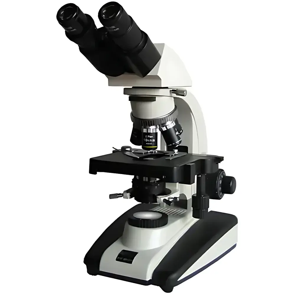


BM XSP-BM-20 UIS Biological Microscope
| Brand | BM |
|---|---|
| Origin | Shanghai, China |
| Microscope Type | Upright Microscope |
| Eyepiece Configuration | Binocular |
| Optical System | UIS Infinity-Corrected |
| Objective Lenses | 4×/0.10, 10×/0.25, 40×/0.65 (Spring), 100×/1.25 (Oil Immersion) |
| Eyepieces | WF10×/22 mm, WF16×/15 mm |
| Total Magnification Range | 40×–1600× |
| Mechanical Stage | Dual-layer, 155 × 145 mm, 80 × 50 mm Travel |
| Focus Mechanism | Coaxial Coarse/Fine Adjustment, 30 mm Travel, Fine Focus Graduation: 2 µm |
| Condenser | Abbe N.A. 1.25 with Centering Adjustment and Iris Diaphragm |
| Illumination | 6 V / 20 W Halogen Lamp, Köhler Illumination, Continuously Variable Brightness Control |
| Filter Set | Blue, Green, Yellow |
| Included Accessories | Immersion Oil, Spare Bulb (6 V / 20 W), Fuse (1 A), Power Cord, User Manual, Certificate of Conformance, Warranty Card |
Overview
The BM XSP-BM-20 UIS Biological Microscope is an upright, infinity-corrected optical instrument engineered for routine and advanced life science applications in academic teaching laboratories, clinical pathology units, and industrial R&D environments. Built upon a UIS (Universal Infinity System) optical architecture, it supports modular expansion—including optional fluorescence modules, phase contrast attachments, and vertical illumination units—enabling seamless adaptation to evolving experimental requirements. Its halogen-based Köhler illumination system ensures uniform, glare-free field illumination across the full magnification range (40× to 1600×), while the fixed 30° inclined, 360° rotatable binocular head accommodates diverse user ergonomics without compromising optical alignment stability. Designed for reproducible quantitative observation, the microscope conforms to fundamental ISO 10934-1 (Microscopy — Vocabulary) and JIS B 7151 (Japanese Industrial Standard for Microscopes) specifications for mechanical and optical performance.
Key Features
- UIS infinity-corrected optical path delivering high-resolution, low-aberration imaging across all objective magnifications
- Four standard plan achromat objectives: 4× (NA 0.10, WD 20.0 mm), 10× (NA 0.25, WD 13.5 mm), 40× (NA 0.65, spring-loaded, WD 0.68 mm), and oil-immersion 100× (NA 1.25, WD 0.20 mm)
- Wide-field eyepieces: 10×/22 mm and 16×/15 mm, both fully corrected for flat-field viewing and optimized for extended visual sessions
- Coaxial coarse/fine focusing mechanism with 30 mm total travel, fine focus graduation of 2 µm, and adjustable coarse-focus tension control
- Dual-layer mechanical stage (155 × 145 mm) with 80 × 50 mm calibrated translation range and vernier scale for precise specimen repositioning
- N.A. 1.25 Abbe condenser with centering screws and iris diaphragm for optimal resolution and contrast control per magnification setting
- 6 V / 20 W halogen lamp with continuous brightness regulation and integrated Köhler alignment optics for consistent illumination intensity and evenness
Sample Compatibility & Compliance
The XSP-BM-20 is compatible with standard 1″ × 3″ (25 × 75 mm) glass slides and 0.17 mm cover slips. It supports transmitted-light modalities only (brightfield, darkfield via optional condenser modification, and basic color-filtered contrast enhancement). While not certified for GLP or GMP-regulated production environments, its mechanical repeatability, standardized optical parameters, and traceable calibration pathways align with ASTM E2877-21 (Standard Guide for Microscope Calibration) and support audit-ready documentation for educational accreditation (e.g., ABET) and institutional biosafety protocols. The instrument meets CE marking requirements for laboratory electrical safety (EN 61010-1) and electromagnetic compatibility (EN 61326-1).
Software & Data Management
The XSP-BM-20 operates as a standalone optical platform and does not include embedded digital imaging hardware. However, it features a standardized trinocular port (optional accessory) enabling integration with C-mount-compatible CMOS/CCD cameras (e.g., 1/2″ or 1/1.8″ sensors). When paired with third-party microscopy software (such as ImageJ, Olympus cellSens, or Thorlabs ThorCam), users can acquire, annotate, measure (length, area, particle count), and export TIFF/PNG images compliant with FAIR data principles. Audit trail functionality, electronic signatures, and 21 CFR Part 11 compliance are achievable only when deployed within validated software ecosystems operated under documented SOPs.
Applications
This microscope serves core functions across multiple domains: undergraduate histology and microbiology labs (e.g., Gram staining analysis, mitotic index quantification); clinical cytology screening (e.g., peripheral blood smear evaluation); pharmaceutical QC (e.g., excipient particle morphology assessment); and agricultural botany (e.g., pollen grain characterization). Its robust mechanical design and thermal-stable illumination make it suitable for prolonged classroom use and multi-user shared-resource facilities. The oil-immersion 100× objective enables reliable visualization of subcellular structures including bacterial flagella, nuclear chromatin patterns, and protozoan organelles—provided specimens are appropriately fixed, stained, and mounted.
FAQ
Is the XSP-BM-20 compatible with fluorescence imaging?
No—fluorescence capability requires a dedicated epi-illumination unit, filter cube turret, and high-sensitivity detector; these are not included and must be added as optional accessories.
What is the maximum usable magnification with the 100× oil objective?
The theoretical maximum useful magnification is ~1250×, limited by visible light wavelength and numerical aperture; higher nominal magnifications (e.g., 1600× with 16× eyepieces) yield empty magnification without additional resolution.
Can the microscope be adapted for photomicrography?
Yes—via optional trinocular head and C-mount adapter, supporting standard 1/2″ or 1/1.8″ sensor cameras with appropriate relay lens configuration.
Does the instrument support phase contrast or differential interference contrast (DIC)?
Phase contrast is possible with a matching phase condenser and phase objectives (not supplied); DIC requires specialized Nomarski prisms and polarizers, which are outside the base configuration.
What maintenance is required for long-term optical performance?
Annual cleaning of optical surfaces using lens-grade tissue and solvent, periodic verification of Köhler alignment, and replacement of the halogen lamp every 100–150 operating hours are recommended practices.


