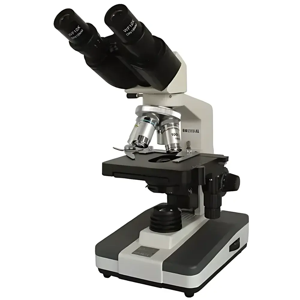

BM XSP-BM-2C Biological Microscope
| Brand | BM |
|---|---|
| Origin | Shanghai, China |
| Microscope Type | Upright |
| Eyepiece Configuration | Binocular |
| Mechanical Tube Length | 160 mm |
| Objective Lenses | 4× (NA 0.10, WD 37.5 mm), 10× (NA 0.25, WD 7.31 mm), 40× (NA 0.65, WD 0.63 mm), 100× oil immersion (NA 1.25, WD 0.18 mm) |
| Eyepieces | Wide-field 10×/Φ18 mm and 16×/Φ11 mm |
| Total Magnification Range | 40×–1600× |
| Stage | Mechanical 130×130 mm with 70×30 mm travel and 0.1 mm vernier scale |
| Interpupillary Adjustment | 55–75 mm |
| Focusing System | Coaxial coarse/fine focus with 15 mm travel |
| fine focus graduation | 0.002 mm |
| Condenser | Abbe condenser, NA 1.25, adjustable iris diaphragm |
| Filters | Blue, green, yellow |
| Illumination | 6 V / 20 W halogen or LED lamp, continuously variable brightness |
Overview
The BM XSP-BM-2C Biological Microscope is an upright, binocular research-grade optical microscope engineered for precision life science applications in academic laboratories, clinical diagnostics, pharmaceutical quality control, and routine histopathology. Designed in accordance with classical finite-conjugate optical architecture (mechanical tube length: 160 mm), it employs achromatic objective lenses and wide-field plan eyepieces to deliver high-fidelity, low-aberration imaging across a total magnification range of 40× to 1600×. Its modular optical path supports both transmitted brightfield illumination and optional phase contrast or darkfield configurations via accessory compatibility. The instrument integrates a robust mechanical stage with calibrated X–Y translation, coaxial focusing with sub-micron fine adjustment resolution (0.002 mm per division), and a high-NA Abbe condenser (N.A. 1.25) optimized for critical resolution at high magnifications—including oil-immersion observation at 100×. The 30° inclined, 360° rotatable binocular head accommodates extended user sessions while maintaining ergonomic alignment.
Key Features
- Upright finite-conjugate optical design compliant with ISO 8578 and JIS B 7131 standards for biological microscopy
- Binocular viewing head with interpupillary adjustment (55–75 mm) and 30° inclination for sustained visual comfort
- Four-parfocal achromatic objectives (4×, 10×, 40×, 100× oil) with standardized RMS threading and engraved numerical aperture (NA) and working distance (WD) specifications
- Wide-field plan eyepieces (10×/Φ18 mm and 16×/Φ11 mm) providing extended field of view and edge-to-edge flatness
- Coaxial coarse/fine focusing mechanism with 15 mm vertical travel and micrometer-scale fine focus (0.002 mm graduation)
- 130 × 130 mm mechanical stage with vernier-calibrated 70 × 30 mm travel and 0.1 mm positional resolution
- Adjustable Abbe condenser (N.A. 1.25) with centering screws and iris diaphragm for Köhler illumination optimization
- Dual illumination options: 6 V / 20 W halogen lamp or energy-efficient LED source, both with continuous brightness control
- Integrated filter holder supporting standardized interference and absorption filters (blue, green, yellow) for contrast enhancement and spectral selection
Sample Compatibility & Compliance
The XSP-BM-2C is validated for use with standard glass microscope slides (76 × 26 mm) and coverslips (No. 1.5, 0.17 mm thickness). It supports aqueous, glycerol, and immersion oil-based mounting media—particularly for high-resolution observation using the 100× oil-immersion objective. The microscope meets general requirements for routine clinical microscopy under CLIA and CAP guidelines when operated within defined environmental conditions (20–25°C, <60% RH). While not certified for GMP-regulated environments out-of-the-box, its optical traceability, mechanical reproducibility, and stable illumination output support integration into GLP-compliant workflows when paired with documented calibration procedures and maintenance logs. All optical components are coated with anti-reflection layers to minimize stray light and maximize contrast transfer.
Software & Data Management
The XSP-BM-2C is a standalone optical instrument without built-in digital imaging or proprietary software. However, it features a standardized trinocular port (optional adapter required) compatible with C-mount or 23.2 mm eyepiece-tube interfaces for third-party CCD/CMOS cameras (e.g., Thorlabs, Lumenera, or Basler models). When integrated with image acquisition platforms such as NIS-Elements (Nikon), ZEN (Zeiss), or open-source tools like ImageJ/Fiji, the system supports quantitative morphometric analysis, multi-channel overlay, time-lapse capture, and export of TIFF/OME-TIFF metadata-rich files. Audit trail functionality, electronic signatures, and 21 CFR Part 11 compliance are achievable through validated laboratory information management systems (LIMS) or digital microscopy suites deployed on networked workstations—provided instrument usage, calibration events, and user access are logged externally.
Applications
- Cell morphology assessment in hematology (e.g., peripheral blood smear analysis per CLSI H20-A2)
- Microbial identification and Gram staining evaluation in clinical microbiology labs
- Plant and animal tissue section examination in histology teaching and research
- Pharmaceutical particle characterization during dissolution testing or excipient inspection
- In vitro cytotoxicity screening and live-cell observation (with appropriate environmental chamber adaptation)
- Quality assurance of medical device coatings and biocompatible material surfaces
- Undergraduate and graduate-level instruction in cell biology, genetics, and pathology curricula
FAQ
Is the XSP-BM-2C compatible with digital camera integration?
Yes—via optional trinocular attachment and standard C-mount or 23.2 mm adapters. Camera selection must match the microscope’s finite conjugate design and sensor size constraints.
Does this microscope support phase contrast or fluorescence?
It does not include phase contrast sliders or fluorescence filter cubes by default, but the optical train permits retrofitting with manufacturer-approved accessories or third-party kits meeting RMS thread and back focal plane specifications.
What is the recommended maintenance schedule?
Daily cleaning of optics with lens tissue and appropriate solvent; quarterly verification of Köhler illumination alignment; annual recalibration of mechanical stage verniers and focus scale accuracy per internal SOPs.
Can the 100× oil-immersion objective be used with non-oil immersion media?
No—using immersion oil is mandatory for optimal resolution and correction at 100×. Substitution with water or glycerol will result in severe spherical aberration and reduced NA utilization.
Is service documentation available in English?
Yes—English-language operation manuals, parts diagrams, and technical bulletins are provided upon request and conform to IEC 61000-6-3 EMC and IEC 61010-1 safety directives.
Related Products


