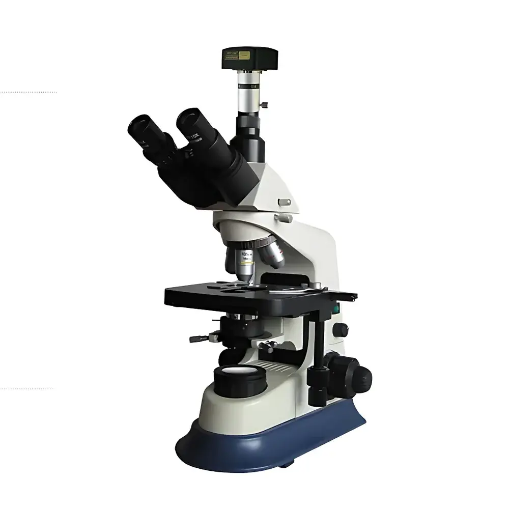


BM XSP-BM-30AD Computer-Enabled Biological Microscope with UIS Optical System
| Brand | BM |
|---|---|
| Origin | Shanghai, China |
| Model | XSP-BM-30AD |
| Microscope Type | Computer-Integrated Biological Microscope |
| Eyepiece Configuration | Binocular |
| Optical System | Infinity-Corrected UIS (Universal Intermediate System) |
| Eyepieces | Widefield Plan 10×/Φ22 mm and 16×/Φ15 mm |
| Objectives | Infinity-Corrected Achromatic Plan Objectives (4×/0.10, 10×/0.25, 40×/0.65, 100× oil/1.25) |
| Total Magnification Range | 40×–1600× |
| Mechanical Stage | 175 × 150 mm with 80 × 50 mm travel and 0.1 mm vernier scale |
| Interpupillary Adjustment | 55–75 mm |
| Focus Mechanism | Coaxial coarse/fine focusing (30 mm travel), fine focus graduation: 0.002 mm, built-in stop and tension control |
| Condenser | Abbe condenser, N.A. 1.25, centerable and iris diaphragm adjustable |
| Filters | Blue, green, yellow interference filters |
| Illumination | 6 V / 20 W halogen lamp with Köhler illumination and continuous brightness control |
| Digital Imaging System | 5 MP CMOS camera with MCL-Z C-mount adapter |
Overview
The BM XSP-BM-30AD is an infinity-corrected, computer-integrated biological microscope engineered for routine and advanced life science applications in academic laboratories, clinical diagnostics, pharmaceutical quality control, and biomedical research. Built upon a Universal Intermediate System (UIS) optical architecture, it delivers high-fidelity contrast, consistent color fidelity, and minimal chromatic aberration across the full magnification range (40× to 1600×). Its Köhler-illuminated halogen light source ensures uniform specimen illumination and optimal resolution at all magnifications, while the rigid mechanical stage and coaxial focusing system provide stable, repeatable positioning—critical for time-lapse imaging and comparative histological analysis. Designed for long-term reliability in shared-core facilities, the instrument conforms to ISO 10934-1 (optical microscopy terminology) and supports GLP-compliant documentation when paired with validated imaging workflows.
Key Features
- Infinity-corrected optical path with plan achromatic objectives (4×, 10×, 40×, 100× oil), enabling modular expansion with fluorescence or phase contrast accessories
- Binocular inclined head (30° tilt, 360° rotation) with interpupillary adjustment (55–75 mm) and dual widefield plan eyepieces (10×/Φ22 mm and 16×/Φ15 mm)
- Precision mechanical stage (175 × 150 mm) with calibrated 80 × 50 mm travel and 0.1 mm vernier readout for reproducible region-of-interest navigation
- Coaxial coarse/fine focusing system with 0.002 mm fine-focus graduation, integrated upper limit stop, and user-adjustable coarse-focus tension
- N.A. 1.25 Abbe condenser with centering mechanism and variable aperture diaphragm—optimized for both brightfield and critical resolution work
- Digital imaging integration via MCL-Z C-mount adapter and 5-megapixel CMOS camera, supporting real-time acquisition and post-acquisition metrology
Sample Compatibility & Compliance
The XSP-BM-30AD accommodates standard 1″ × 3″ glass slides and 24 × 50 mm coverslips, compatible with paraffin-embedded tissue sections, cytology smears, live-cell monolayers (on chambered coverslips), and stained microbiological preparations. Its optical design meets ASTM E2877-22 (Standard Guide for Microscope Calibration), and its illumination stability complies with ISO 9241-307 (Ergonomics of human-system interaction — Part 307: Requirements for electronic visual displays used in microscopy). While not intrinsically FDA-listed, the system supports 21 CFR Part 11–compliant data handling when used with validated third-party image management software and audit-trail-enabled acquisition protocols. It is routinely deployed in ISO/IEC 17025-accredited testing labs for morphological QC of raw materials and finished biopharmaceutical products.
Software & Data Management
The included geometric measurement software provides calibrated, traceable quantification of linear dimensions, angular relationships, circularity, area, and profile deviations. All measurements are referenced to user-defined stage micrometer calibrations and support export of annotated TIFF/PNG images with embedded scale bars and metadata (date, objective, magnification, camera settings). Raw image data is stored in lossless formats; no proprietary compression is applied. The software does not require cloud connectivity and operates offline—ensuring compliance with institutional data sovereignty policies. For integration into LIMS or ELN environments, CSV and XML exports are supported, and the API allows scripted batch processing of multi-field montages.
Applications
- Routine hematology and urinalysis (blood smear differential counts, cast identification, crystal morphology)
- Histopathological screening of H&E- and IHC-stained tissue sections in teaching and diagnostic labs
- Microbial colony morphology assessment and Gram-stain interpretation in QC microbiology
- Cell culture monitoring—including confluence estimation, mitotic index scoring, and contamination detection
- Pharmaceutical excipient particle sizing and distribution analysis per USP
- Botanical and mycological specimen documentation requiring high-resolution structural annotation
FAQ
Is the XSP-BM-30AD compatible with fluorescence imaging?
The base configuration supports brightfield only. Fluorescence capability requires optional filter cubes, excitation/emission filters, and a dedicated epi-illuminator—not included in standard delivery.
Can the 5 MP camera be upgraded to higher resolution?
Yes—BM offers validated 3 MP and 10 MP CMOS modules with identical C-mount interface and software driver compatibility.
Does the microscope meet CE or UL safety standards?
The instrument complies with IEC 61010-1:2010 for laboratory electrical equipment safety and carries the CE marking for the European Economic Area.
What maintenance intervals are recommended for the halogen illumination system?
Lamp replacement is advised every 100–150 hours of cumulative use; condenser alignment and objective cleaning should occur quarterly in high-throughput environments.
Is remote operation or networked image streaming supported?
Native network streaming is not implemented; however, third-party VNC or LabVIEW-based DAQ integration enables supervised remote acquisition within secured LAN environments.


