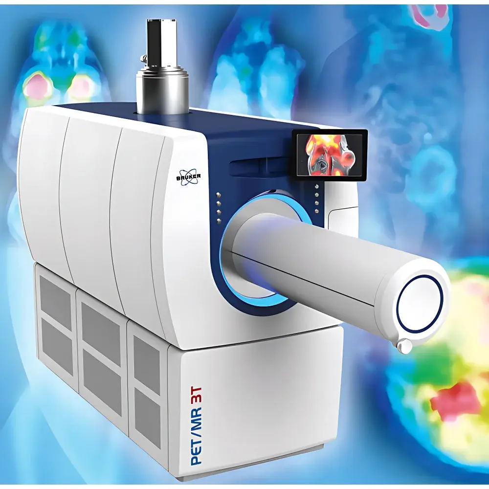
Bruker PET/MR 3T Small Animal In Vivo Imaging System
| Brand | Bruker |
|---|---|
| Origin | USA |
| Manufacturer Type | Authorized Distributor |
| Origin Category | Imported |
| Model | PET/MR 3T |
| Imaging Modality | Integrated Positron Emission Tomography / Magnetic Resonance Imaging |
| Energy Resolution | 17% FWHM at 511 keV |
| Spatial Resolution (PET) | <0.7 mm (NEMA NU-4) |
| Spatial Resolution (MRI) | ≤1.2 mm isotropic (with CryoProbe™) |
| Scan Speed | Up to 10 min per dynamic PET frame (variable by protocol) |
| Field of View (FOV) | 80 mm (PET transaxial), 148 mm (MRI transaxial), 285 mm (whole-body MRI longitudinal) |
| Animal Capacity | 1 subject per scan (mouse or rat) |
Overview
The Bruker PET/MR 3T Small Animal In Vivo Imaging System is a fully integrated, preclinical dual-modality platform combining high-field 3 Tesla magnetic resonance imaging (MRI) with time-of-flight (TOF) positron emission tomography (PET). Engineered for precision and reproducibility in longitudinal animal studies, it operates on the principle of simultaneous data acquisition—where PET detects metabolic and molecular tracer distribution via gamma-ray coincidence detection, while MRI provides anatomical context, soft-tissue contrast, and functional parameters (e.g., diffusion, perfusion, BOLD) without ionizing radiation. Unlike sequential or co-registered systems, this platform achieves true temporal and spatial synchronization: PET detectors are embedded within the MRI magnet bore, enabling concurrent acquisition with minimal mutual interference. The system utilizes a novel helium-free 3T superconducting magnet, eliminating cryogen dependency while maintaining field homogeneity (<0.1 ppm over 40 mm DSV) and thermal stability essential for quantitative MRI. Its compact architecture supports standardized housing in dedicated small-animal imaging suites compliant with IACUC and AAALAC facility requirements.
Key Features
- Simultaneous PET/MRI acquisition with hardware-synchronized timing (<100 ns jitter), ensuring voxel-wise correlation between molecular signal and anatomical/functional MRI contrast.
- Sub-millimeter PET spatial resolution: 0.7 mm full-width-at-half-maximum (FWHM) at center-of-FOV, validated per NEMA NU-4 standards—enabling reliable quantification in murine brain substructures and orthotopic tumor models.
- High-sensitivity PET detector ring incorporating lutetium-yttrium oxyorthosilicate (LYSO) crystals and silicon photomultipliers (SiPMs), delivering 12% system sensitivity (at center, 25 mm radius) for reduced radiotracer dose and shorter scan durations.
- Integrated MRI CryoProbe™ technology: 4-channel transmit/receive cryogenic RF coil optimized for mice and rats, providing ≥2× SNR gain over room-temperature coils—critical for high-resolution T2-weighted, DTI, and fMRI protocols.
- Motorized, touch-screen-controlled animal handling system with real-time position feedback, enabling repeatable stereotactic positioning across sessions and automatic coregistration of multi-session datasets.
- Zero-boil-off 3T magnet with active shielding and passive shimming—reducing siting footprint, operational overhead, and long-term maintenance costs versus conventional liquid-helium-dependent systems.
Sample Compatibility & Compliance
The system accommodates live, anesthetized rodents (C57BL/6 mice, Sprague-Dawley rats) in standardized physiological monitoring setups (respiratory gating, temperature control, ECG triggering). It complies with ISO 10993-1 (biocompatibility of animal interface components), IEC 61000-6-3 (EMC for laboratory equipment), and supports GLP-compliant workflows through audit-trail-enabled acquisition logs. All PET reconstruction algorithms adhere to NEMA NU-4 and NU-2 standards; MRI sequences are validated against ACR MRI phantom test protocols. Data export formats (DICOM-RT, NIfTI, MINC) ensure interoperability with institutional PACS and regulatory submission packages (e.g., FDA IND-enabling toxicology imaging).
Software & Data Management
Acquisition and reconstruction are managed via ParaVision® 360, Bruker’s clinical-preclinical imaging platform supporting automated pulse sequence programming, real-time motion correction, and vendor-neutral DICOM export. PET data undergoes iterative OSEM reconstruction with MR-based attenuation correction (MRAC) using Dixon-segmented tissue classification—eliminating CT-derived attenuation maps and associated radiation exposure. Quantitative analysis leverages PMOD v4.3+ with kinetic modeling (e.g., Logan plot, Patlak analysis), ROI-based SUV normalization, and voxel-wise parametric mapping. Audit trails, user authentication, and electronic signatures comply with 21 CFR Part 11 requirements when deployed in regulated environments. Raw data archives are stored in Bruker’s proprietary format (FID/Bruker) with lossless compression and SHA-256 checksum verification.
Applications
- Oncology: Longitudinal tracking of [¹⁸F]FDG uptake in orthotopic glioblastoma or [⁶⁴Cu]DOTATATE binding in neuroendocrine tumors—correlated with MRI-derived tumor volume, necrosis fraction, and permeability (Ktrans).
- Neuroscience: Simultaneous assessment of amyloid-β deposition ([¹¹C]PiB PET) and hippocampal microstructural integrity (DTI + T2* mapping) in Alzheimer’s disease models.
- Cardiology: Dual-gated PET/MRI for myocardial glucose metabolism ([¹⁸F]FDG) synchronized with cine-MRI-derived ejection fraction and strain analysis in ischemia-reperfusion models.
- Inflammation & Immunology: [⁸⁹Zr]oxine-labeled T-cell trafficking imaged alongside MRI-based edema mapping in EAE-induced demyelination.
- Pharmacokinetics: Real-time visualization of radiolabeled nanocarrier biodistribution coupled with dynamic contrast-enhanced (DCE) MRI to quantify vascular permeability in tumor stroma.
FAQ
Is the PET/MR 3T compatible with standard radiotracers used in preclinical research?
Yes—the system supports all common cyclotron-produced tracers ([¹⁸F], [⁶⁴Cu], [⁸⁹Zr], [¹¹C]) and generator-based isotopes ([⁶⁸Ga], [⁹⁹mTc]), with energy windowing optimized for 511 keV gamma detection.
How is attenuation correction performed without CT?
MR-based attenuation correction (MRAC) uses a dual-echo Dixon MRI sequence to segment tissue into fat, water, lung, and background—assigning appropriate linear attenuation coefficients for PET quantification.
Can the system perform whole-body scans in a single session?
Yes—motorized animal handling enables sequential bed-positioning across three FOVs (80 mm PET, 148 mm MRI, up to 285 mm longitudinal MRI), with automatic stitching and intensity normalization in ParaVision.
What level of technical support and service coverage is available internationally?
Bruker offers global service contracts including preventive maintenance, remote diagnostics, on-site engineer response (<48 h), and software update licensing—aligned with ISO 9001-certified service operations.
Does the system support retrospective motion correction?
Yes—retrospective PET motion correction is enabled via optical tracking markers integrated into the animal bed, synchronized with MRI navigator echoes for respiratory and cardiac motion compensation.
Related Products


