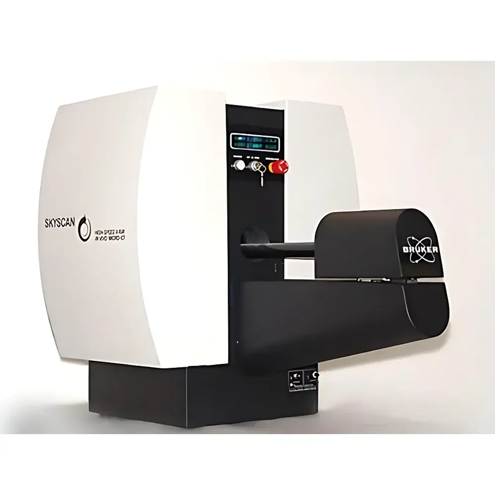
Bruker SKYSCAN 1178 High-Throughput In Vivo Micro-CT Imaging System
| Brand | Bruker |
|---|---|
| Origin | Germany |
| Manufacturer Type | Authorized Distributor |
| Origin Category | Imported |
| Model | SKYSCAN 1178 |
| Instrument Type | Tomographic Imaging |
| X-ray Source | Sealed Metal-Ceramic Tube (20–65 kV, 40 W) |
| Detector | Digital X-ray Camera (1280 × 1024 pixels, 12-bit) |
| Scan Volume | Ø82 mm × 82 mm (single scan), up to 200 mm full-length |
| Voxel Size (isotropic) | 80 µm (1024³) or 160 µm (512³) |
| Minimum Scan Time | <45 s (160 µm) |
| Full-body mouse scan | 3 min 20 s (80 µm) |
| Reconstruction Time | 45 s (160 µm) |
| Animal Handling | Carbon-fiber mouse/rat bed on interchangeable mounting platform |
| Physiological Monitoring | Real-time respiration & ECG gating, motion detection |
| Radiation Safety | Surface dose rate <1 µSv/h during operation |
| Software Suite | NRecon, DataViewer, CTAn, CTVol, MorphoGraphX |
Overview
The Bruker SKYSCAN 1178 is a high-throughput, in vivo micro-computed tomography (micro-CT) imaging system engineered for longitudinal preclinical research in small animal models. Operating on the principle of cone-beam X-ray computed tomography, it acquires projection data from multiple angular positions and reconstructs high-fidelity 3D volumetric datasets using filtered back-projection and iterative algorithms. Designed specifically for routine, quantitative in vivo studies, the system delivers isotropic spatial resolution down to 80 µm with sub-minute volumetric reconstruction—enabling rapid screening of skeletal architecture, lung morphology, tumor burden, vascular calcification, and implant integration without sacrificing structural fidelity. Its integrated physiological monitoring and respiratory/ECG gating capability ensure motion artifact suppression in conscious or lightly sedated animals, supporting GLP-compliant longitudinal study designs across oncology, musculoskeletal, cardiovascular, and pulmonary disease models.
Key Features
- Sub-45-second whole-body mouse scan at 160 µm isotropic resolution; full-resolution (80 µm) whole-body acquisition completed in under 3 minutes 20 seconds
- Real-time cluster-based reconstruction: image volumes generated concurrently with data acquisition, reducing total workflow latency to under 60 seconds
- Dedicated carbon-fiber animal bed with modular, interchangeable mounting interface—compatible with multimodal integration platforms for PET, SPECT, and bioluminescence imaging
- Integrated physiological monitoring subsystem: non-invasive respiration belt, ECG electrodes, and motion detection sensors provide synchronized gating signals for motion-compensated acquisition
- Sealed metal-ceramic X-ray source (20–65 kV, 40 W) paired with a high-dynamic-range digital X-ray detector (1280 × 1024, 12-bit) ensures stable beam output and low-noise projection data
- Compliance with IEC 61000-6-3 (EMC) and IEC 62471 (photobiological safety); surface radiation exposure maintained below 1 µSv/h during operation per DIN 6812
Sample Compatibility & Compliance
The SKYSCAN 1178 accommodates live mice and rats (up to ~500 g) in standardized positioning protocols optimized for reproducible thoracic, abdominal, and skeletal region-of-interest targeting. The carbon-fiber bed minimizes X-ray attenuation while providing mechanical stability under anesthesia or light sedation. All hardware and software components conform to ISO 13485:2016 (medical device quality management) and support audit-ready documentation required for FDA 21 CFR Part 11 compliance when deployed in regulated environments. Image metadata—including acquisition parameters, calibration timestamps, and user authentication logs—are embedded and preserved throughout the processing pipeline, fulfilling ALARA (As Low As Reasonably Achievable) dosimetry reporting and GLP traceability requirements.
Software & Data Management
The bundled SkyScan software suite provides end-to-end workflow automation: NRecon performs GPU-accelerated reconstruction with ring artifact correction and beam hardening compensation; DataViewer enables slice navigation and orthogonal reformatting; CTAn delivers ISO/ASTM-standardized bone morphometry (BV/TV, Tb.Th, Tb.Sp, Conn.D), tissue density quantification (HU calibration), and region-growing segmentation; CTVol generates publication-grade volume renderings with adjustable opacity mapping and surface smoothing. All modules support batch processing, scripting via Python API, and DICOM export compliant with PACS integration standards (DICOM Supplement 113). Audit trails record operator actions, parameter changes, and version-controlled analysis pipelines—essential for regulatory submissions under USP , ISO/IEC 17025, and OECD GLP Principles.
Applications
- Longitudinal bone phenotyping: trabecular and cortical bone remodeling in osteoporosis, arthritis, and metastatic bone disease models
- Pulmonary structure-function correlation: emphysema progression, fibrosis staging, and aerosol deposition mapping
- Oncology: primary tumor volume kinetics, metastatic lesion enumeration, and orthotopic model validation
- Cardiovascular research: aortic calcification scoring, stent deployment assessment, and myocardial infarction modeling
- Biomaterial evaluation: scaffold degradation kinetics, osseointegration metrics, and vascular ingrowth quantification
- Multimodal correlation: co-registration of CT-derived anatomy with PET tracer uptake or bioluminescent signal distribution
FAQ
What is the minimum achievable voxel size for in vivo scanning?
The system achieves isotropic voxels as small as 80 µm (1024³ matrix) under standard in vivo acquisition conditions. Sub-50 µm resolution is technically feasible ex vivo but not recommended for live-animal studies due to increased radiation dose and prolonged scan time.
Can the SKYSCAN 1178 be integrated into an existing multimodal imaging facility?
Yes—the interchangeable animal bed platform supports mechanical and electrical interfacing with Bruker’s Albira PET/SPECT systems, IVIS Spectrum CT, and third-party optical imaging stations via standardized mounting rails and TTL-synchronized trigger I/O.
Does the system meet regulatory requirements for preclinical contract research organizations (CROs)?
All software modules include electronic signature support, audit trail logging, and configurable user access levels aligned with 21 CFR Part 11 and EU Annex 11 expectations for data integrity in GLP-compliant studies.
How is radiation dose managed during repeated in vivo scans?
The system implements automatic kV/mA optimization, beam filtration presets, and dose-tracking reports per scan session. Dose maps are exported alongside reconstructed volumes to enable cumulative exposure monitoring across longitudinal cohorts.
Is remote operation and cluster reconstruction supported?
Yes—reconstruction nodes can be distributed across a local network; acquisition control and real-time preview are accessible via secure RDP or web-based ThinLinc client, enabling off-site experiment monitoring without compromising data security.


