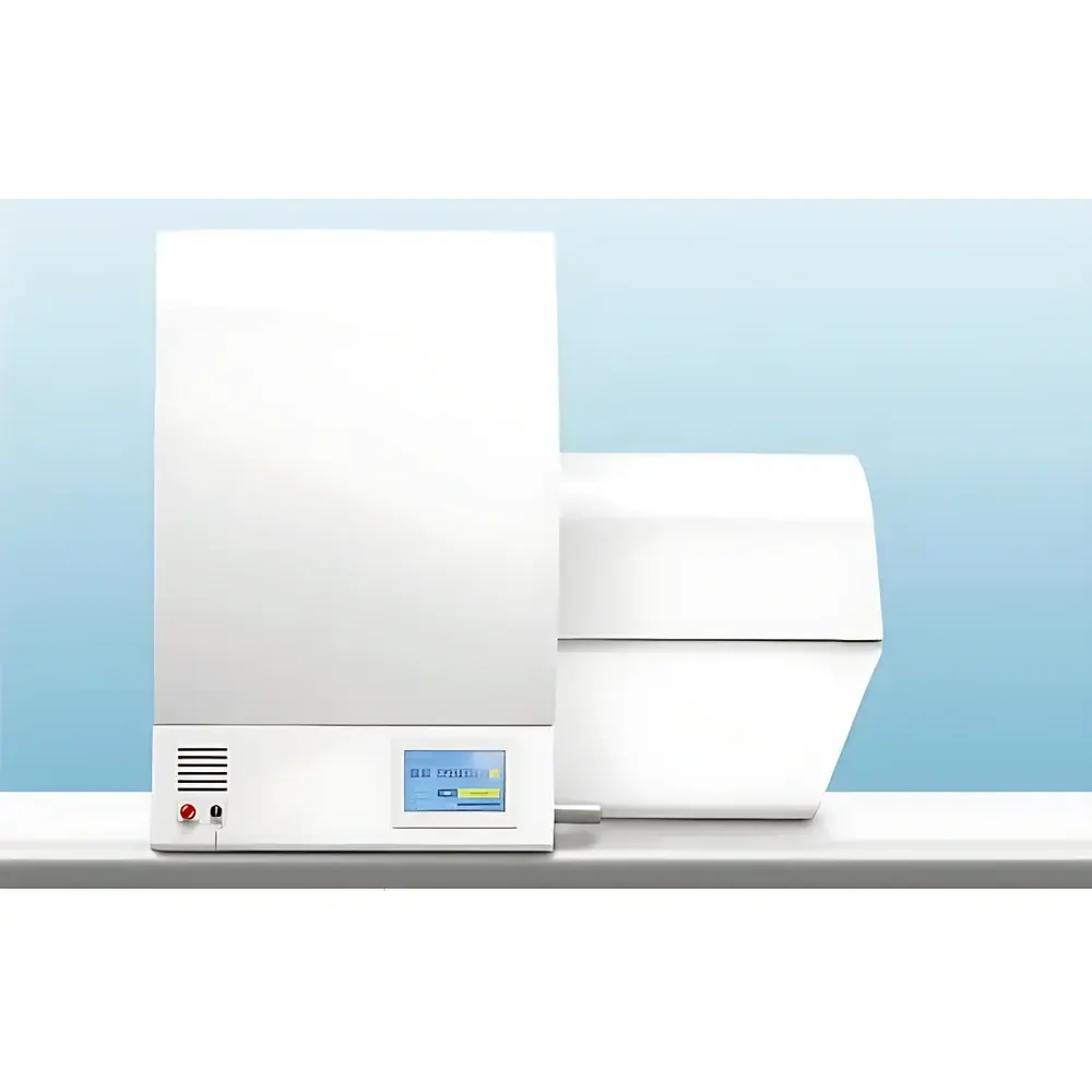
Bruker SkyScan 1276 In Vivo Micro-CT Imaging System
| Brand | Bruker |
|---|---|
| Origin | Germany |
| Model | SkyScan 1276 |
| Imaging Modality | X-ray micro-computed tomography (micro-CT) |
| Spatial Resolution (nominal) | ≤2.8 µm |
| Scan Modes | Step-and-shoot, continuous gantry rotation with slip-ring interface |
| Scan Time per Reconstruction | as low as 3.9 s |
| Reconstructed Slice Count per Scan | >1600 slices |
| Max Image Matrix Size | 8000 × 8000 pixels |
| X-ray Source Voltage Range | 20–100 kV adjustable |
| Automatic Filter Changer | 6-position |
| Dose Reduction Technology | Beam-shaping collimator (patented by Bruker microCT) |
| Physiological Monitoring Integration | Respiratory gating, ECG synchronization, temperature stabilization, motion detection |
| Animal Handling | Interchangeable mouse/rat beds with integrated anesthesia mask and sensor interface |
| Real-time Dose Monitoring | On-screen dosimetry derived from projection image analysis |
| Reconstruction Acceleration | GPU-accelerated algorithms + InstaRecon® layered reconstruction (≥10× faster than conventional FBP) |
| Software Compliance | GLP-compliant software suite with audit trail, user access control, and electronic signature support |
| Data Export & Remote Viewing | Direct export to iOS/Android devices via dedicated viewer app |
Overview
The Bruker SkyScan 1276 is a high-performance, benchtop in vivo micro-computed tomography (micro-CT) system engineered for longitudinal, non-invasive 3D structural imaging of small laboratory animals—primarily mice and rats—under physiological conditions. Operating on the principle of cone-beam X-ray tomography, the system acquires hundreds to thousands of angular projections during controlled gantry rotation, followed by iterative or filtered back-projection (FBP)-based reconstruction into isotropic volumetric datasets. Its core architecture integrates a high-stability microfocus X-ray source, a large-area flat-panel detector, and a precision motorized sample stage with integrated physiological monitoring interfaces. Designed specifically for preclinical research environments, the SkyScan 1276 delivers sub-3 µm nominal spatial resolution while maintaining dose efficiency critical for repeated imaging studies. The system supports both static and dynamic (4D) acquisition protocols, enabling time-resolved morphological assessment synchronized to cardiac and respiratory cycles—essential for cardiovascular, pulmonary, oncology, and bone metabolism investigations.
Key Features
- Continuous variable geometric magnification with nominal spatial resolution down to 2.8 µm, achieved through precise source-detector-object distance optimization.
- Slip-ring-enabled continuous gantry rotation eliminates mechanical stop-start limitations, supporting both step-and-shoot and true helical scanning modes.
- Ultra-fast acquisition capability: full-volume scans completed in as little as 3.9 seconds per reconstructed tomogram, with up to 1600 slices per scan at up to 8000 × 8000 pixel matrix size.
- Bruker-patented beam-shaping collimator reduces absorbed radiation dose by 2–3× without compromising contrast-to-noise ratio or spatial fidelity.
- Integrated physiological monitoring suite—including respiratory pressure sensor, ECG electrodes, thermal regulation module, and motion tracking—enables robust 4D gated reconstruction.
- Modular animal handling platform with quick-swap mouse/rat beds, integrated isoflurane anesthesia delivery, and unified cabling for all biosignal inputs.
- Real-time on-screen dosimetry calculates and displays estimated effective dose per scan based on projection intensity, tube voltage, current, and filtration—supporting ALARA compliance.
Sample Compatibility & Compliance
The SkyScan 1276 accommodates live rodents within standardized, ventilated animal beds equipped with gas anesthesia ports and embedded sensor connectors. Optional accessories extend compatibility to ex vivo tissue samples, biopsies, and biomaterial scaffolds. All hardware and firmware comply with IEC 61000-6-3 (EMC), IEC 62471 (photobiological safety), and EU Directive 2013/59/Euratom for ionizing radiation protection. The system’s software package adheres to Good Laboratory Practice (GLP) requirements per OECD Series on Principles of GLP, including full audit trail logging, role-based user authentication, electronic signatures (in accordance with FDA 21 CFR Part 11), and data integrity safeguards throughout acquisition, reconstruction, and analysis workflows.
Software & Data Management
NRecon® reconstruction software provides GPU-accelerated iterative and FBP algorithms, while InstaRecon®—Bruker’s proprietary layered reconstruction engine—delivers ≥10× speed improvement over conventional methods without loss of quantitative accuracy. CTAn® enables automated segmentation, morphometric analysis (e.g., BV/TV, Tb.Th, Conn.D), and statistical reporting with batch-processing support. Data export is natively compatible with DICOM 3.0, NIfTI, and TIFF formats. The optional mobile viewer application allows secure, real-time remote visualization of reconstructed volumes on iOS and Android tablets—ideal for collaborative review and presentation. All software modules operate under a centralized license manager with version-controlled updates and documented change history.
Applications
- Longitudinal bone phenotyping: trabecular architecture quantification, osteoporosis progression monitoring, and fracture healing assessment.
- Cardiovascular research: coronary artery calcification scoring, left ventricular mass/volume dynamics, and myocardial infarction modeling.
- Oncology: tumor volume kinetics, angiogenesis evaluation via contrast-enhanced perfusion imaging, and metastatic burden quantification.
- Pulmonary studies: airway remodeling, emphysema grading, and functional ventilation mapping using dual-energy or xenon-enhanced protocols.
- Developmental biology: embryonic skeletal maturation, craniofacial morphogenesis, and organ volumetry across gestational stages.
- Biomaterial integration: scaffold degradation profiling, host-tissue ingrowth analysis, and mechanical property correlation via density mapping.
FAQ
What is the minimum achievable voxel size in vivo?
The system achieves a nominal isotropic voxel size of 2.8 µm under optimal geometric magnification and high-resolution acquisition settings—subject to animal size, positioning stability, and motion gating efficacy.
Can the SkyScan 1276 perform contrast-enhanced imaging?
Yes—when used with iodinated or lanthanide-based contrast agents and appropriate kVp/filtration selection, the system supports soft-tissue differentiation and vascular perfusion mapping.
Is the system compliant with FDA 21 CFR Part 11 for regulated studies?
The GLP software package includes electronic signature capability, audit trail generation, and user access controls aligned with 21 CFR Part 11 requirements for nonclinical laboratory studies.
How is radiation dose monitored and reported during scanning?
A real-time dosimeter algorithm analyzes raw projection images to estimate entrance dose and effective dose per scan, displaying results directly on the operator touchscreen interface.
Does the system support multi-modal data fusion?
Yes—CTAn and DataViewer modules support co-registration with PET, SPECT, or optical imaging datasets via common coordinate frameworks and landmark-based alignment tools.


