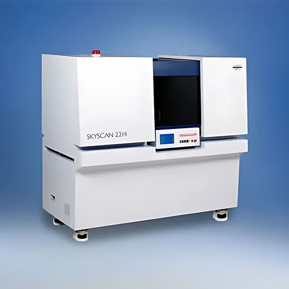
Bruker SKYSCAN 2214 Nanoscale X-ray Micro-CT System
| Brand | Bruker |
|---|---|
| Origin | Belgium |
| Model | SKYSCAN 2214 |
| X-ray Source | 20–100 kV, 10 W, <5 µm focal spot size at 4 W |
| Maximum Reconstruction Volume | 14456 × 14456 × 2630 px (16 MP detector) or 11840 × 11840 × 2150 px (11 MP detector) |
| Spatial Resolution | <0.35 µm (16 MP), <0.45 µm (11 MP) |
| Sample Capacity | Ø ≤ 75 mm, H ≤ 70 mm |
| Detector | 16 MP (4904 × 3280 px) or 11 MP (4032 × 2688 px), 14-bit cooled CCD coupled 1:1 to scintillator via fiber optic taper |
| Density Resolution | <0.35 µm (16 MP), <0.45 µm (11 MP) |
Overview
The Bruker SKYSCAN 2214 is a high-performance nanoscale X-ray micro-computed tomography (micro-CT) system engineered for non-destructive, quantitative 3D imaging across materials science, geoscience, life sciences, and industrial R&D. Operating on the principle of cone-beam X-ray absorption tomography, the system acquires hundreds to thousands of projection images as the sample rotates through 360°, enabling reconstruction of volumetric datasets with true isotropic spatial resolution down to <0.35 µm. Its dual-detector architecture—supporting both 16 MP and 11 MP configurations—and ultra-stable high-brightness microfocus X-ray source with diamond transmission window (<500 nm effective focal spot) ensure exceptional signal-to-noise ratio and geometric fidelity. Designed for laboratory integration rather than cleanroom dependency, the SKYSCAN 2214 delivers sub-micron metrology-grade dimensional accuracy without requiring synchrotron access, making it suitable for routine QA/QC, failure analysis, and longitudinal in situ studies.
Key Features
- Sub-500 nm effective focal spot X-ray source with diamond window, enabling high-resolution imaging of low-contrast and dense samples
- Modular detector platform supporting up to four interchangeable detectors—including optional high-dynamic-range and fast-frame-rate variants—for field-upgradable performance expansion
- InstaRecon® real-time GPU-accelerated 3D reconstruction engine, reducing typical 1000-projection reconstructions from hours to minutes while preserving quantitative fidelity
- Proprietary helical scanning algorithm for continuous sample translation during rotation, eliminating step-and-repeat artifacts and extending effective field-of-view beyond physical detector limits
- Thermally stabilized mechanical stage with active drift compensation, ensuring sub-pixel registration stability over multi-hour acquisitions
- Integrated environmental chamber compatibility (optional) for in situ heating, cooling, compression, or fluid saturation experiments under controlled conditions
Sample Compatibility & Compliance
The SKYSCAN 2214 accommodates a broad range of specimen geometries and densities—from soft biological tissues and polymer foams to metal alloys and geological core plugs—within its maximum sample envelope (Ø75 mm × H70 mm). Its flexible mounting interface supports custom fixtures, tensile stages, and flow cells compliant with ASTM E1441 (Standard Practice for Computed Tomography), ISO 12799 (Micro-CT for Materials Characterization), and USP (Imaging Techniques for Pharmaceutical Dosage Forms). All acquisition and reconstruction workflows are traceable and audit-ready, supporting GLP/GMP environments through configurable electronic logbooks, user authentication, and 21 CFR Part 11–compliant data integrity controls when deployed with Bruker’s CTAn/CTVol software suite.
Software & Data Management
Acquisition, reconstruction, segmentation, and quantification are unified within Bruker’s CTAn (CT Analyzer) and CTVol (CT Volume) software platform. CTAn provides ISO 16232-compliant pore network analysis, grain size distribution (ASTM E112), phase fraction quantification, and directional anisotropy metrics using adaptive thresholding and machine learning–assisted region-growing algorithms. CTVol enables volume rendering, cross-sectional slicing, and export of geometry-ready STL or voxel-based NRRD files for finite element modeling (FEM) or additive manufacturing validation. Raw projections and reconstructed volumes are stored in DICOM-compliant format with embedded metadata (kV, µA, exposure time, magnification, filter settings), ensuring interoperability with PACS systems and third-party analysis tools such as Avizo or Dragonfly.
Applications
- Geosciences: Quantitative characterization of pore-throat networks, mineral phase distribution (e.g., quartz, clay, pyrite), and permeability prediction in reservoir rock cores; time-resolved imaging of fluid displacement during imbibition or drainage
- Advanced Materials: Fiber orientation mapping in carbon-fiber composites, void content analysis in additively manufactured metals, and thermal barrier coating delamination assessment
- Battery Research: In situ 3D monitoring of electrode cracking, lithium plating, separator deformation, and cathode particle fracture during electrochemical cycling
- Life Sciences: Morphometric analysis of trabecular bone architecture (BV/TV, Tb.Th, Tb.Sp), vascular network topology in tumor xenografts, and dental enamel microstructure without staining or sectioning
- Electronics & Packaging: Solder joint void detection, wire bond integrity verification, and encapsulant delamination mapping in IC packages
FAQ
What is the minimum achievable voxel size under standard operating conditions?
The system achieves <0.35 µm isotropic voxel resolution with the 16 MP detector configuration at optimal magnification and exposure settings, validated using NIST-traceable line-pair test objects.
Can the SKYSCAN 2214 perform dynamic in situ experiments?
Yes—when integrated with compatible mechanical, thermal, or electrochemical stages, the system supports time-lapse tomography with temporal resolution down to ~30 seconds per full-volume reconstruction using partial-angle acquisition strategies.
Is helical scanning supported natively, and what are its advantages?
Helical scanning is fully implemented in firmware and software, enabling seamless long-sample imaging (e.g., drill cores or battery cells) with improved Z-axis resolution uniformity and reduced ring artifact incidence compared to conventional step-and-shoot protocols.
How does the system ensure measurement traceability and regulatory compliance?
All hardware parameters are logged with timestamps and user IDs; calibration routines follow ISO/IEC 17025 guidelines; and software modules support 21 CFR Part 11 electronic signatures, audit trails, and role-based access control for regulated laboratories.
What detector upgrade paths are available post-purchase?
The modular detector bay allows field replacement or addition of higher-resolution, faster-readout, or low-noise detectors—including future-generation sensors—without system requalification or major recalibration.


