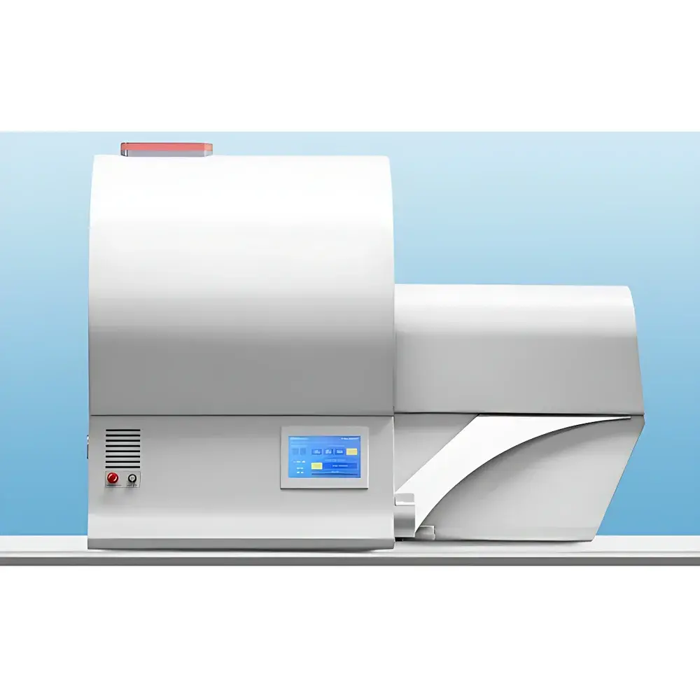
Bruker SkyScan1278 In Vivo Micro-CT Imaging System
| Brand | Bruker |
|---|---|
| Origin | Germany |
| Manufacturer Type | Authorized Distributor |
| Origin Category | Imported |
| Model | SkyScan1278 |
| Instrument Type | Tomographic Imaging System |
| Energy Resolution | 17% |
| Spatial Resolution | 50 µm |
| Scan Time | 7.2 s per full-body mouse scan |
| Field of View Diameter | 80 mm |
| Scan Length | 200 mm |
| Sample Capacity | 1 animal per scan |
| X-ray Source | Adjustable 20–65 kV microfocus tube with patented beam shaping optics |
| Detector | High-sensitivity CMOS flat-panel detector |
| Dose Level | <6 mGy for whole-body murine scan |
| Physiological Gating | Integrated respiratory, ECG, temperature, and motion monitoring |
| Data Output Formats | DICOM, TIFF, JPG, BMP, PNG, AVI |
| Software Compliance | GLP-compliant acquisition and reconstruction suite |
Overview
The Bruker SkyScan1278 is a preclinical in vivo micro-computed tomography (micro-CT) system engineered for high-speed, low-dose, high-fidelity volumetric imaging of small laboratory animals—primarily mice and rats—under physiological conditions. Operating on the principle of cone-beam X-ray tomography, the system acquires projection data from multiple angular positions and reconstructs them into isotropic 3D voxel datasets using filtered back-projection or iterative algorithms. Its core design prioritizes temporal resolution without compromising spatial fidelity: a complete whole-body murine scan is acquired in just 7.2 seconds, enabling dynamic assessment of organ function—including cardiac and pulmonary motion—without exogenous contrast agents. The system integrates a microfocus X-ray source (20–65 kV), a high-gain CMOS flat-panel detector, and Bruker’s proprietary spatial beam shaper, which reduces absorbed radiation dose by a factor of 2–5 while preserving signal-to-noise ratio and quantitative accuracy. This makes the SkyScan1278 uniquely suited for longitudinal studies requiring repeated imaging sessions under strict ALP (Animal Laboratory Practice) and IACUC compliance.
Key Features
- Ultra-fast acquisition: Full-body mouse scan completed in 7.2 seconds at sub-6 mGy effective dose—among the fastest in-class in vivo micro-CT systems.
- Isotropic spatial resolution of 50 µm at the center of rotation, supporting detailed morphometric analysis of bone microarchitecture, vasculature, and soft-tissue interfaces.
- Patented beam-shaping optics that modulate X-ray fluence distribution to minimize peripheral dose while maintaining central image uniformity and quantitative linearity.
- Integrated physiological gating platform: hardware-synchronized acquisition triggered by real-time respiratory waveform, ECG R-wave detection, thermal feedback, and motion tracking—enabling true 4D (3D + time) functional imaging.
- Touchscreen-based operator interface designed for glove-compatible interaction; all critical acquisition parameters—including kV, current, filter selection, and gating thresholds—are adjustable directly from the console.
- Onboard real-time radiation dosimetry: dose calculation engine estimates and displays cumulative absorbed dose per scan based on exposure parameters, geometry, and attenuation maps—supporting ethical dose optimization per ICRP and AAALAC guidelines.
Sample Compatibility & Compliance
The SkyScan1278 accommodates live rodents up to 30 g (mouse) or 200 g (rat) within its 80 mm diameter field of view and 200 mm axial scan length. An integrated gas anesthesia delivery module—compatible with isoflurane and oxygen mixtures—is housed in a dedicated animal transfer chamber with temperature-regulated bedding and CO₂ scrubbing. All physiological monitoring modules (respiratory belt, ECG electrodes, rectal thermistor, and motion sensor) connect via standardized analog/digital I/O ports and are fully software-controlled through the acquisition interface. The system meets ISO 13485 design control requirements for research-use-only (RUO) medical devices and supports Good Laboratory Practice (GLP) workflows via audit-trail-enabled software logging, user access controls, electronic signatures, and versioned protocol archiving—fully aligned with OECD GLP principles and FDA expectations for nonclinical study data integrity.
Software & Data Management
Acquisition, reconstruction, and analysis are unified within Bruker’s NRecon and CTAn software suite. Raw projections are reconstructed into DICOM-compliant volumetric stacks (with embedded metadata including dose, geometry, and gating timestamps), ensuring seamless integration with PACS, third-party visualization tools (e.g., Amira, Avizo), and AI-based segmentation pipelines. Quantitative outputs—including bone volume fraction (BV/TV), trabecular thickness (Tb.Th), cortical porosity, lung density histograms, and cardiac ejection fraction—are traceable to NIST-traceable phantoms and validated against ASTM E1695 and ISO 15708 standards. All processed data can be exported in TIFF, PNG, JPG, BMP, or AVI formats; reconstructed volumes are also accessible via mobile-optimized web viewer for remote review on iOS and Android platforms. Data provenance is preserved through immutable acquisition logs, parameter snapshots, and user-action timestamps compliant with 21 CFR Part 11 Annex 11 requirements.
Applications
The SkyScan1278 serves as a core platform across multiple preclinical domains: longitudinal osteoporosis and bone metastasis modeling; pulmonary structural phenotyping in COPD, fibrosis, and asthma models; cardiac functional assessment in myocardial infarction and hypertrophy studies; oncology (orthotopic tumor volume, angiogenesis, calcification); and developmental biology (embryonic skeletal patterning). Its low-dose capability enables serial imaging over weeks or months—critical for therapeutic efficacy evaluation and safety pharmacology. The absence of iodinated contrast requirements expands utility in renal-impaired models and eliminates confounding hemodynamic effects during functional assessment.
FAQ
What is the minimum detectable feature size in vivo?
The system achieves an isotropic voxel size of 50 µm under standard whole-body murine scanning conditions; smaller features may be resolved in targeted high-resolution scans (e.g., femoral head or calvaria) using magnified geometry.
Can the system perform retrospective gating?
No—gating is strictly prospective and hardware-triggered; respiratory and ECG signals are acquired synchronously with projection acquisition to ensure phase-consistent reconstruction.
Is dose monitoring compliant with regulatory reporting standards?
Yes—the integrated dosimetry module reports absorbed dose in mGy per scan with uncertainty estimation, conforming to ICRP Publication 103 and EU Directive 2013/59/Euratom requirements for justification and optimization.
Does the software support automated segmentation of soft tissues?
CTAn includes semi-automated thresholding, region-growing, and machine learning–assisted segmentation modules; custom AI models can be deployed via Python API integration.
How is system calibration maintained between scans?
Daily air-scan normalization and periodic phantom-based QC (using hydroxyapatite and polyethylene inserts) are enforced by the software’s built-in QA workflow, with results logged and exportable for audit.


