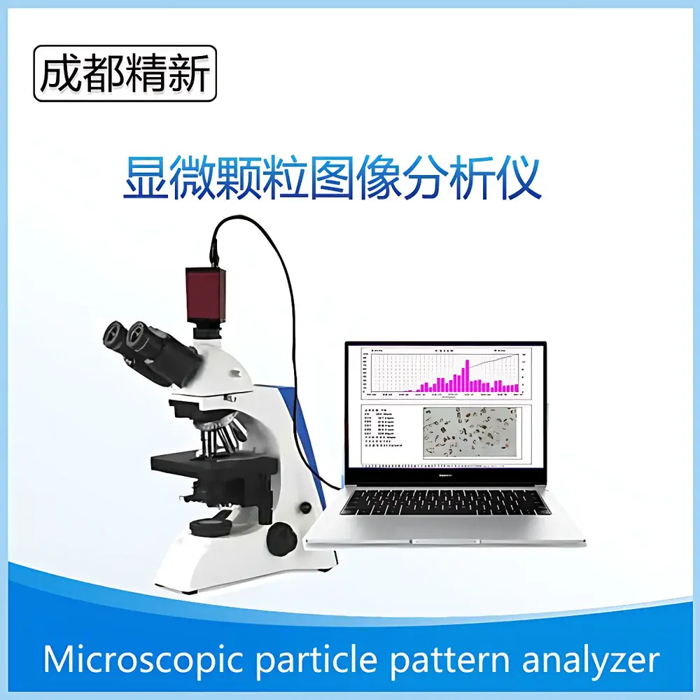
Chengdu Jingxin JX-2000A Microscopic Particle Image Analyzer
| Brand | Chengdu Jingxin (CDJX) |
|---|---|
| Origin | Sichuan, China |
| Manufacturer Type | Direct Manufacturer |
| Instrument Type | Optical Particle Counter |
| Model | JX-2000A |
| Measurement Range | 0.5–3000 µm |
| Max. Optical Magnification | 1600× |
| Max. Total Magnification (Digital + Optical) | ≤4000× |
| Resolution | ≤0.1 µm |
| Microscope Configuration | Transmitted-light Microscope (optional configurations: domestic, metallurgical, or imported) |
| CCD Camera | 3 MP or 5 MP |
| Eyepieces | 10×, 16× |
| Objectives | 4×, 10×, 20×, 40×, 100× |
| Calibration Scale | 10 µm |
| Software Platform | Windows 7 / Windows 10 |
| Interface | USB 2.0 High-Speed Data Transfer |
| Analytical Parameters | Particle Size Distribution, Aspect Ratio Distribution, Circularity Distribution, D10/D50/D90/D97, Mean Diameter, Surface Area, Particle Count, Area/Perimeter/Volume Metrics |
Overview
The Chengdu Jingxin JX-2000A Microscopic Particle Image Analyzer is a high-resolution optical particle characterization system engineered for quantitative morphological and dimensional analysis of dispersed solid particles in powders, slurries, and suspensions. Unlike ensemble-averaging techniques such as laser diffraction, this instrument employs direct digital imaging via transmitted-light microscopy combined with advanced image segmentation and geometric feature extraction algorithms. It captures true two-dimensional projections of individual particles, enabling precise measurement of size distribution, shape descriptors—including circularity, aspect ratio, convexity, and solidity—and surface texture attributes. The system operates on the principle of calibrated pixel-to-length conversion, where each image is referenced against a certified micrometer scale (10 µm), ensuring traceable dimensional metrology compliant with ISO 13322-1:2020 (Particle size analysis — Image analysis methods — Part 1: Static image analysis). Designed for laboratories requiring orthogonal validation of laser diffraction or dynamic light scattering data, the JX-2000A serves as a reference-grade tool for method verification, process development, and quality control in regulated environments.
Key Features
- Modular optical platform supporting interchangeable microscope configurations: standard transmitted-light, optional metallurgical (reflected-light), or imported high-NA objectives for enhanced contrast and resolution
- Dual-resolution CCD imaging options (3 MP or 5 MP) with real-time preview, low-noise analog-to-digital conversion, and adjustable exposure/gain controls for optimal contrast across diverse sample refractive indices
- Comprehensive digital zoom and geometric transformation suite: 90° rotations (clockwise/counterclockwise), horizontal/vertical mirroring, arbitrary scaling, and sub-pixel interpolation for accurate spatial registration
- Multi-stage image enhancement pipeline including histogram equalization, morphological operations (erosion/dilation), edge-preserving smoothing, adaptive thresholding, and binary mask refinement
- Calibration-flexible measurement engine supporting unit switching between µm, mm, cm, and inch—critical for cross-scale reporting and regulatory documentation
- Batch processing mode enabling sequential analysis of ≥50 images per session, with automatic stitching of statistical aggregates to improve sampling representativeness and reduce measurement uncertainty
- Customizable analysis templates for industry-specific materials (e.g., abrasive grains, lithium cobalt oxide cathode powders, mica flakes), configurable via XML-based protocol definitions
Sample Compatibility & Compliance
The JX-2000A accommodates dry powders, liquid dispersions (aqueous/organic), and semi-solid pastes when mounted on standard glass slides or graticule slides. Sample preparation follows ISO 13322-1 Annex B guidelines: controlled deposition, uniform monolayer formation, and refractive index matching where applicable. Validated applications span mineral fillers (barium sulfate, quartz, silicon carbide), battery electrode materials (LiCoO₂, graphite), pharmaceutical excipients (microcrystalline cellulose, lactose), catalyst supports (alumina, silica), and functional pigments. All analytical outputs—including D10/D50/D90, circularity histograms, and particle count density—are exportable in CSV and PDF formats compatible with LIMS integration. While not inherently 21 CFR Part 11–compliant out-of-the-box, audit trail functionality (user log, timestamped parameter changes, versioned report generation) can be enabled via optional software modules meeting GLP/GMP documentation requirements.
Software & Data Management
The proprietary Jingxin Image Analysis Suite runs natively on Windows 7/10 (64-bit) and communicates with hardware via USB 2.0 with deterministic latency (<15 ms frame capture interval). Raw image metadata (exposure time, gain, objective ID, calibration factor) is embedded in EXIF headers. Processed results include annotated overlays showing Feret diameter, minimum bounding rectangle, convex hull, and centroid coordinates. Statistical summaries are generated per ISO 9276-2:2014 (Representation of results of particle size analysis — Part 2: Calculation of average particle sizes/diameters and moments from particle size distributions). Reports support customizable headers (sample ID, operator, date/time, dispersion medium, sonication duration), embedded thumbnail galleries, and dual-language (English/Chinese) export—essential for multinational QC workflows. Data integrity is preserved through SHA-256 checksums applied to exported datasets.
Applications
- Orthogonal verification of laser diffraction (ISO 13320) and sedimentation (ISO 13317) results in R&D and release testing
- Morphology-driven formulation optimization: correlating aspect ratio distribution with flowability (ASTM D6393) or compaction behavior
- FDA-regulated pharmaceutical powder characterization per USP particulate matter testing guidance
- Quality gate monitoring for abrasive grain grading (FEPA 43-2019) and catalyst pellet uniformity assessment
- Failure analysis of agglomerated or sintered particles in battery slurry manufacturing
- Educational use in materials science curricula for hands-on particle metrology training
FAQ
What is the smallest detectable particle size under optimal conditions?
With 100× objective and 5 MP CCD, the theoretical resolution limit is ≤0.1 µm; however, reliable detection requires ≥3–5 pixels per particle dimension, yielding a practical lower bound of ~0.5 µm for spherical, high-contrast particles in monolayer preparations.
Can the system analyze transparent or low-contrast particles like fused silica or PMMA?
Yes—via optional dark-field illumination kits or refractive index-matching media (e.g., Cargille immersion oils), contrast can be significantly enhanced without altering particle morphology.
Is third-party software integration supported?
Raw TIFF/Bitmap exports and structured CSV outputs enable seamless import into MATLAB, Python (OpenCV/scikit-image), or commercial platforms such as ImageJ/Fiji and MATLAB Particle Analyzer Toolboxes.
How is calibration traceability maintained?
Each system ships with NIST-traceable stage micrometers (10 µm pitch); calibration routines validate pixel-to-length mapping at every objective magnification and are logged with operator signature and timestamp.
Does the software support automated focus stacking?
No—focus stacking requires manual Z-axis adjustment per field of view; however, the software allows multi-plane image registration and composite overlay for qualitative depth assessment.
Related Products


