Chengyi RM6240S Physiological Signal & Video Teaching Acquisition System
| Brand | Chengyi (SCChengyi) |
|---|---|
| Origin | Sichuan, China |
| Model | RM6240S |
| Video Sensor | 1/3" CCD, 2 MP |
| Optical Zoom | ≥12× |
| Digital Zoom | ≥8× |
| Max Composite Magnification | 105× |
| Video Resolution (Digital/USB) | 2048×1024 @ 15 fps |
| Analog Output | VGA/AV, 800 TV lines @ 30 fps |
| SNR | ≥48 dB |
| Sync Latency (Video vs. Physio Signal) | <0.1 s |
| Lighting | Dual LED side illumination + cold-cathode backlight (≥2 W) |
| Input/Output | Dual VGA, Dual RGB (DB15), AV (RCA), RS-232, Audio In/Out |
| Software | USB video capture module + synchronized physiological waveform recording (MPEG-4 compression), standalone or integrated mode, annotation, freeze, invert, zoom, rotation, text overlay |
Overview
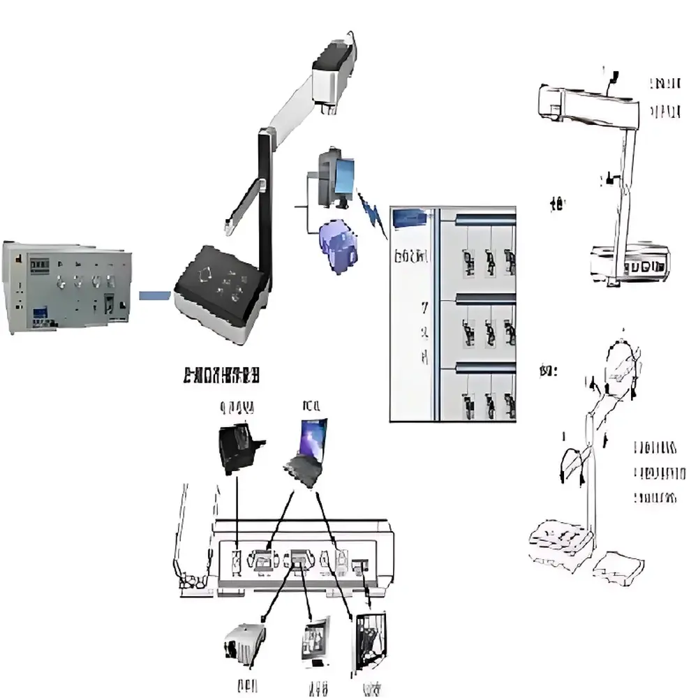
The Chengyi RM6240S Physiological Signal & Video Teaching Acquisition System is an integrated hardware-software platform engineered for high-fidelity, time-synchronized acquisition and pedagogical presentation of live physiological experiments. Designed explicitly for undergraduate and graduate biomedical education laboratories, it bridges the gap between real-time electrophysiological or biomechanical signal acquisition and visual demonstration by embedding a calibrated video channel directly into the data acquisition workflow. The system operates on a dual-stream architecture: one analog/digital channel captures physiological waveforms (e.g., ECG, EMG, arterial pressure, nerve conduction) from compatible multi-channel acquisition units (e.g., RM6240BD/CD), while a synchronized HD video stream—captured via a motorized, optically stabilized 1/3″ CCD camera—records surgical procedures, tissue preparations, or instrument manipulation. Temporal alignment is maintained with sub-100 ms latency (<0.1 s), ensuring waveform events (e.g., stimulus onset, spike latency, contractile peak) correspond precisely to visual context—a requirement for rigorous teaching assessment and reproducible lab reporting.
Key Features
- Real-time synchronization of physiological signals and HD video: Integrated software enables concurrent acquisition, display, playback, and export of both data streams with timestamp-aligned metadata.
- Motorized optical platform: Compact foldable design (270 × 185 × 40 mm collapsed; max height 465 mm extended) with 330° pan capability and dual-axis camera articulation for optimal specimen framing.
- High-fidelity imaging subsystem: 12× optical zoom + 8× digital zoom (105× composite), auto-focus, auto-iris, and manual/automatic white balance; supports both image and text modes for enhanced legibility during slide or document projection.
- Dual illumination architecture: Front-facing LED side lamps and rear-mounted cold-cathode backlight (≥2 W) ensure uniform, shadow-free illumination across biological specimens, surgical tools, and printed materials—without heat-induced tissue degradation.
- Flexible I/O interface suite: Dual VGA inputs/outputs, dual RGB (DB15), AV (RCA), RS-232 serial control, stereo audio in/out, enabling seamless integration with projectors, interactive whiteboards, recording PCs, and legacy lab equipment.
- Hardware-accelerated video processing: Onboard functions include color-to-grayscale conversion, frame freeze, positive/negative inversion, brightness/contrast/saturation adjustment, geometric scaling, and rotational correction—accessible via front-panel buttons or IR remote control.
Sample Compatibility & Compliance
The RM6240S accommodates diverse instructional use cases—including isolated organ bath recordings, neuromuscular preparations, small-animal surgery demonstrations, and medical device operation training. Its non-contact optical design eliminates contamination risk during sterile procedures, and its low-heat LED lighting preserves tissue viability during prolonged observation. While not certified for clinical diagnostic use, the system complies with general-purpose educational laboratory safety standards (IEC 61010-1). Data export formats—including MPEG-4 compressed video (.mp4), annotated screenshots (.png/.jpg), and time-stamped waveform files (.txt/.mat)—support interoperability with LMS platforms (e.g., Moodle, Canvas) and institutional archival systems. All software modules adhere to standard Windows OS security protocols and permit audit-trail–enabled user session logging when deployed in GLP-aligned academic settings.
Software & Data Management
The bundled acquisition software provides two operational modes: standalone video capture (for lecture preparation or independent documentation) and integrated synchronous recording (with RM6240BD/CD or equivalent physiological signal processors). Video is acquired at up to 2048 × 1024 resolution via USB 2.0 or at 800 TV lines via analog VGA/AV input. To preserve temporal fidelity during long-duration experiments, users may select lower-resolution recording profiles (320 × 240 or 640 × 480) without aspect-ratio distortion. All video is encoded using MPEG-4 compression to minimize storage footprint while retaining diagnostic-grade clarity. Annotation tools support dynamic text overlays, freehand drawing, region-of-interest magnification, and frame-by-frame navigation. Exported video files are codec-standard (.mp4) and playable in VLC, Windows Media Player, or MATLAB; waveform-video synchronization is preserved in native .chd format and recoverable in third-party analysis environments via CSV timestamp mapping.
Applications
- Undergraduate physiology labs: Visual reinforcement of action potential propagation, synaptic transmission, and cardiovascular dynamics alongside real-time signal traces.
- Surgical skills training: Step-by-step documentation of microdissection, cannulation, or electrode placement with correlated electrophysiological feedback.
- Curriculum development: Creation of reusable, timestamped multimedia lab modules compliant with ABET or AAMC competency frameworks.
- Remote learning infrastructure: Stream-ready output supports hybrid delivery via Zoom, Teams, or dedicated streaming encoders (via HDMI/VGA loop-through).
- Research methodology instruction: Demonstration of proper signal grounding, noise mitigation, and artifact identification through synchronized visual-audio-contextual cues.
FAQ
Can the RM6240S operate independently of the RM6240BD/CD physiological recorder?
Yes—the system includes a standalone USB video capture application that functions without connection to any external signal acquisition hardware.
What video formats does the software support for export?
Primary export format is MPEG-4 (.mp4); still frames may be saved as PNG or JPEG; synchronized waveform-video composites are stored in proprietary .chd format with CSV-compatible timestamps.
Is the video latency compensated during playback?
No active latency compensation is applied; however, hardware-level sync ensures inherent <0.1 s drift between video frames and waveform samples, eliminating perceptible desynchronization during didactic review.
Does the system support FDA 21 CFR Part 11 compliance?
It is not validated for regulated clinical use; however, optional user authentication, session logging, and immutable file export features can be configured to align with institutional GLP/GMP training documentation requirements.
How is calibration handled for quantitative image analysis?
The platform does not include built-in pixel-to-metric calibration; users must apply external reference scales (e.g., stage micrometers) and perform post-hoc spatial normalization in third-party image analysis software (e.g., ImageJ, MATLAB).
Related Products

")