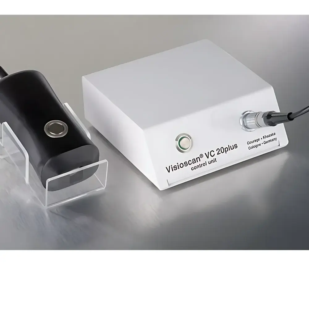
CK VC20plus Skin Surface Texture Analysis System
| Brand | CK |
|---|---|
| Origin | Germany |
| Manufacturer Status | Authorized Distributor |
| Origin Category | Imported |
| Model | VC20plus |
| Pricing | Upon Request |
Overview
The CK VC20plus Skin Surface Texture Analysis System is a non-invasive, high-resolution imaging platform engineered for quantitative assessment of epidermal topography under standardized ultraviolet (UV) illumination. It operates on the principle of UV-induced surface contrast enhancement: keratinized stratum corneum layers fluoresce or reflect UV light differentially compared to underlying viable epidermis, enabling precise grayscale differentiation—where wrinkles and microfurrows appear as low-intensity (dark) regions, while desquamated corneocytes manifest as high-intensity (bright) features. Captured at native optical resolution without contact or mechanical pressure, the system delivers 8-bit grayscale images (256 intensity levels per pixel), forming the basis for objective, operator-independent morphometric evaluation of skin surface architecture. Designed for integration into dermatological research, cosmetic clinical trials, and pharmaceutical development workflows, the VC20plus meets foundational requirements for reproducible, longitudinal skin surface monitoring in compliance with ISO 13842:2021 (Guidance on instrumental methods for assessing skin surface topography) and supports GLP-aligned study documentation.
Key Features
- UV-optimized optical path with narrowband 365 nm excitation source and calibrated filter set to maximize contrast between hydrated and desquamated stratum corneum layers
- Fixed-focus macro lens system delivering consistent field-of-view (12 × 12 mm) and depth-of-field suitable for facial and volar forearm sites
- Integrated LED-based UV illumination ensuring stable irradiance output (< ±2% variation over 10,000 cycles) and eliminating thermal drift during sequential acquisitions
- Real-time image preview with adjustable exposure control to accommodate variable skin phototypes (Fitzpatrick I–VI)
- Embedded hardware synchronization for shutter-illumination timing, minimizing motion artifacts in uncooperative subjects or dynamic assessments
- Rugged aluminum housing rated IP52 for controlled clinical environment use; CE-marked per Directive 2016/425 (PPE) and 2014/30/EU (EMC)
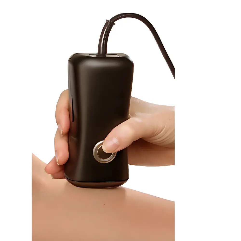
Sample Compatibility & Compliance
The VC20plus is validated for in vivo application on human skin across anatomical sites including forehead, crow’s feet, cheek, dorsal hand, and volar forearm. It requires no coupling gels, occlusion, or pre-treatment—enabling immediate post-application assessment in efficacy studies. All image acquisition protocols align with ISO/IEC 17025:2017 general requirements for competence of testing and calibration laboratories, and data outputs are compatible with audit trails required under FDA 21 CFR Part 11 when paired with CK’s certified software suite. The system complies with IEC 62471:2006 (Photobiological safety of lamps and lamp systems), classifying its UV output as Risk Group 1 (exempt) for short-term exposure under standard operating conditions.
Software & Data Management
Acquisition and analysis are performed using CK’s proprietary Skin Image Analysis Software (v5.2+), which provides automated region-of-interest (ROI) selection, edge-detection-based wrinkle segmentation, and ISO 4287-compliant roughness parameter calculation—including Sa (arithmetic mean height), Sq (root-mean-square height), and Sdr (developed interfacial area ratio). Raw TIFF images retain full 16-bit linear intensity metadata; processed results export to CSV, PDF, and XML formats with embedded timestamps, operator ID, device serial number, and acquisition parameters. Audit log functionality records all user actions—including ROI modifications, threshold adjustments, and report generation—with immutable timestamps, satisfying traceability mandates in GCP, GMP, and cosmetic regulatory submissions (e.g., EU Cosmetics Regulation (EC) No 1223/2009 Annex I technical dossier requirements).
Applications
- Cosmetic ingredient and formulation efficacy testing: quantification of anti-wrinkle, moisturizing, exfoliating, and barrier-repair claims per CTFA/ISO 19779 guidelines
- Dermatopharmacology: objective monitoring of topical corticosteroid-induced atrophy, retinoid-induced desquamation, or calcineurin inhibitor–mediated texture normalization
- Clinical dermatology: baseline documentation and progression tracking in ichthyosis, atopic dermatitis, psoriasis, and photoaging cohorts
- Medical device evaluation: surface interaction assessment of microneedling rollers, radiofrequency devices, and fractional laser resurfacing outcomes
- Regulatory dossier support: generation of instrumentally derived endpoints accepted by EFSA, Health Canada, and ASEAN Cosmetic Committee for substantiation of structural claims
FAQ
Does the VC20plus require skin contact or application of coupling agents?
No—the system is fully non-contact and relies solely on reflected UV light; no gels, powders, or adhesives are used.
Can it be used on darker skin phototypes (Fitzpatrick V–VI)?
Yes—exposure compensation algorithms and UV-specific contrast optimization ensure diagnostic-grade image fidelity across all six Fitzpatrick types.
Is the UV source compliant with occupational safety standards?
Yes—the 365 nm LED array emits below the ACGIH TLV® threshold for chronic UV-A exposure (1.0 J/cm² per 8-hour period); no protective eyewear is mandated beyond standard clinical lighting protocols.
How does VC20plus differ from confocal microscopy or profilometry-based skin analyzers?
Unlike point-scanning or mechanical contact methods, VC20plus captures full-field surface morphology in a single exposure, eliminating registration errors and enabling rapid bilateral or multi-site comparisons without repositioning.
Is raw image data export supported for third-party statistical analysis?
Yes—TIFF files include embedded EXIF metadata (exposure time, gain, lens focal length, UV intensity calibration factor), supporting independent validation and meta-analysis in MATLAB, Python (scikit-image), or R environments.
Related Products