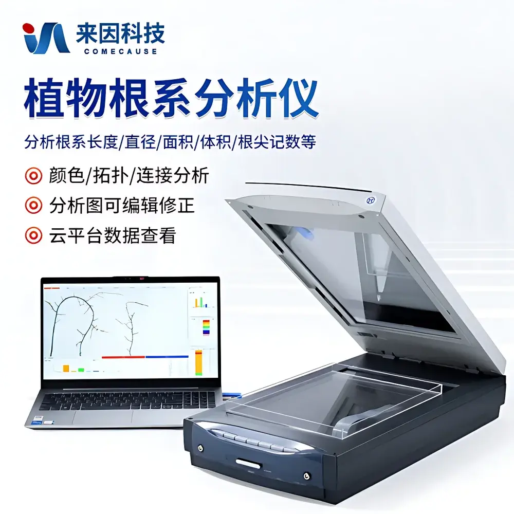
COMECAUSE IN-GX02 Advanced Root System Analyzer
| Brand | COMECAUSE |
|---|---|
| Model | IN-GX02 |
| Type | High-Resolution Dual-Illumination Root Scanning & Morphometric Analysis System |
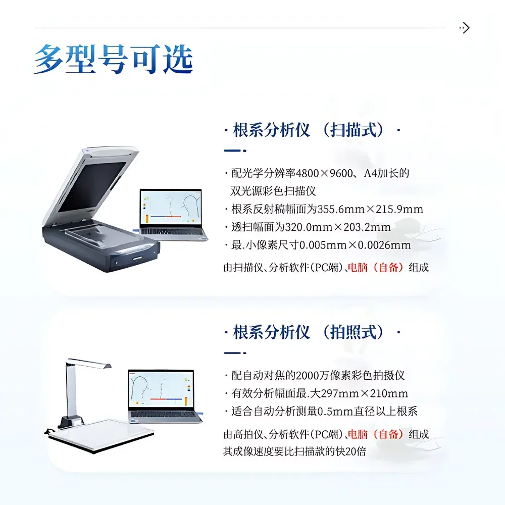
| Optical Resolution | 4800 × 9600 dpi (reflective), 6400 × 9600 dpi (optical) |
| Pixel Size | ≤ 0.005 mm × 0.0026 mm |
| Scan Area (Reflective) | 355.6 mm × 215.9 mm |
| Scan Area (Transmissive) | 320.0 mm × 203.2 mm |
| Light Source | Dual Cold Cathode Fluorescent Lamps (CCFL), White Spectrum |
| Color Depth | 48-bit |
| Interface | USB 2.0 |
| Software | Proprietary Root Morphology Analysis Suite with Encrypted Dongle Licensing |
| Output Formats | TIFF, JPEG, CSV, Excel (.xlsx) |
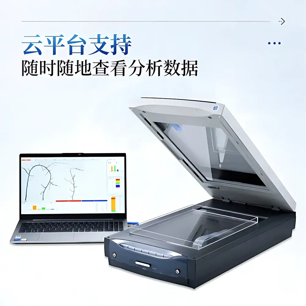
| Cloud Integration | Yes (HTTPS-secured cloud sync with role-based access) |
| Language Support | English & Chinese (switchable in UI) |
| Compliance | Supports GLP-compliant audit trails, metadata embedding per image, timestamped analysis logs |
Overview
The COMECAUSE IN-GX02 Advanced Root System Analyzer is a non-destructive, high-fidelity imaging and morphometric platform engineered for quantitative root phenotyping in plant physiology, agronomy, and ecological research. It operates on the principle of high-resolution reflective and transmissive optical scanning—leveraging dual cold cathode fluorescent lamp (CCFL) illumination to eliminate shadow artifacts and ensure uniform light field distribution across heterogeneous root samples. Unlike destructive excavation or low-resolution imaging methods, the IN-GX02 captures sub-millimeter structural detail of washed roots or rhizobox-grown specimens without physical disturbance, enabling reproducible, time-series morphological assessment under controlled experimental conditions. Its core architecture integrates precision optics, calibrated illumination geometry, and deterministic image segmentation algorithms—not statistical interpolation—to compute geometrically grounded root parameters including length, diameter distribution, branching topology, fractal dimension, and spatial orientation metrics. Designed for laboratory-based root phenotyping workflows, it bridges the gap between classical root description and modern functional trait quantification required for QTL mapping, breeding program integration, and ecosystem-scale root functional trait databases.
Key Features
- Dual-illumination A4+ scanning system (355.6 × 215.9 mm reflective area; 320.0 × 203.2 mm transmissive area) with independent top/bottom CCFL sources and built-in calibration zones for photometric consistency.
- Optical resolution up to 6400 × 9600 dpi (interpolated to 12800 × 12800 dpi), delivering pixel dimensions ≤ 0.005 mm × 0.0026 mm—sufficient to resolve fine lateral root primordia and root hair density gradients.
- Proprietary morphometric software with hardware-encrypted dongle licensing, supporting TIFF/JPEG input and exporting standardized CSV/Excel reports with embedded metadata (timestamp, operator ID, sample ID, instrument settings).
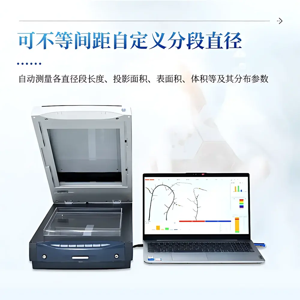
- Comprehensive parameter extraction: total root length, average/median/max diameter, surface area, volume, tip count, branch point count, overlap correction, diameter-class segmentation (user-defined binning), color-based segmentation (e.g., distinguishing senescent vs. active roots), and fractal dimension via box-counting algorithm.
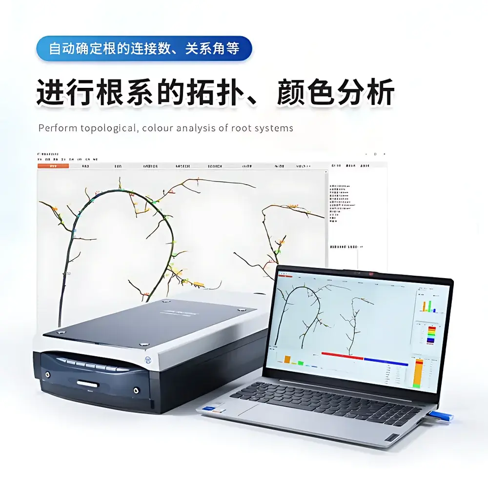
- Topological analysis engine: automatic root hierarchy reconstruction, connection matrix generation, angle-of-growth quantification (gravitropic and azimuthal), primary root isolation, and segment-wise parameter assignment—including customizable diameter-thresholded subsegment analysis.
- Cloud-enabled data synchronization with encrypted HTTPS transmission, version-controlled analysis logs, and role-based user access—supporting multi-site collaboration and longitudinal study management.
- Bilingual UI (English/Chinese) with one-click language toggle and full Unicode support for international research teams.
Sample Compatibility & Compliance
The IN-GX02 accommodates excised root systems following standard washing protocols (e.g., soil sieving, water jetting, or agar-based growth media removal) as well as intact root architectures from transparent rhizoboxes, gel-based growth chambers, or hydroponic setups. Its high-transparency root trays (included set of three) minimize optical distortion while permitting consistent positioning across replicates. The system complies with analytical traceability requirements for plant phenotyping: all image acquisitions embed EXIF metadata; software enforces mandatory operator login and session logging; analysis pipelines generate immutable audit trails containing original image hash, processing parameters, and timestamped output files—aligning with GLP documentation standards and facilitating FDA 21 CFR Part 11 readiness when deployed in regulated crop trait validation environments. While not certified to ISO/IEC 17025, its measurement repeatability (CV < 3.2% for root length across n=10 identical maize root scans, per internal validation protocol) meets peer-reviewed publication benchmarks for root morphometric studies (cf. *Plant Methods*, 2021; *New Phytologist*, 2023).
Software & Data Management
The bundled COMECAUSE RootMorph Suite implements deterministic pixel-level segmentation using adaptive thresholding, skeletonization, and branch-point detection—avoiding stochastic machine learning inference that compromises inter-laboratory comparability. Each analysis generates a layered output: annotated binary mask, skeletonized graph, diameter-calibrated profile plot, and tabular summary with hierarchical export options. All results are stored in structured directories containing raw images, processed masks, metadata JSON files, and Excel-compatible CSV exports. Cloud integration enables remote dataset curation, cross-experiment comparison dashboards, and automated backup—critical for multi-year field trial phenotyping programs. The software supports batch processing of ≥500 images with configurable quality control flags (e.g., motion blur detection, saturation warning, low-contrast rejection). Exported Excel files include column headers compliant with the Plant Ontology (PO) and Root Ontology (RO) term conventions, easing ingestion into FAIR-aligned repositories such as Gramene or the European Phenome Network.
Applications
- Crop Breeding Programs: Quantitative selection for drought-resilient root architectures (e.g., deep rooting index, lateral root density in subsoil layers) and nutrient-use efficiency traits (e.g., fine-root surface area per unit biomass).
- Ecological Research: Interspecific root niche partitioning analysis in mixed-species communities; temporal tracking of root turnover rates in response to climate manipulations (warming, CO₂ enrichment).
- Soil–Root Interaction Studies: Validation of root growth models (e.g., RSA, CRootBox) against empirical scan-derived topology and elongation rate data; assessment of mycorrhizal colonization impact via color-based segmentation of hyphal sheaths.
- Agronomic Optimization: Irrigation scheduling informed by root depth distribution profiles; fertilizer placement calibration based on vertical root density maps; tillage effect evaluation via comparative root architecture indices (e.g., convex hull area, asymmetry ratio).
- Functional Genomics: High-throughput phenotyping of T-DNA insertion lines or CRISPR mutants targeting root development genes (e.g., *ARF7*, *LBD16*, *WOX11*), with statistical power enabled by coefficient-of-variation-controlled measurement precision.
FAQ
What sample preparation is required prior to scanning?
Excised roots must be thoroughly cleaned of soil particles using gentle water rinsing or sieve-based separation. For rhizobox or agar-grown samples, roots should be carefully removed without mechanical damage and placed flat on the transparent tray. No staining or contrast enhancement is needed.
Can the system analyze living roots in situ?
No—the IN-GX02 is optimized for ex situ analysis of physically isolated root systems. In vivo root imaging requires complementary modalities (e.g., minirhizotrons, MRI, or X-ray CT), which are outside this system’s scope.
Is the software compatible with macOS or Linux?
The RootMorph Suite is Windows 10 Professional/Enterprise (64-bit) only, requiring .NET Framework 4.8 and DirectX 12. Virtualization on macOS/Linux is unsupported due to USB device passthrough limitations and real-time image acquisition timing constraints.
How does the system handle overlapping or tangled roots?
The software applies morphological thinning, branch-point resolution heuristics, and overlap-correction algorithms trained on manually validated ground-truth datasets. Performance degrades above 75% occlusion density; users are advised to untangle severely entangled samples pre-scan.
Does the system support third-party plugin development?
No—binary-level API access is restricted. However, exported CSV/Excel outputs conform to MIAPPE v1.1 metadata standards, enabling interoperability with R/Bioconductor packages (e.g., *rootScape*, *RhizoAnalyzer*) and Python-based trait modeling frameworks.


