
COMECAUSE IN-GX02 Advanced Root System Analyzer for High-Resolution Morphometric and Topological Phenotyping
| Brand | COMECAUSE |
|---|---|
| Model | IN-GX02 |
| Origin | Shandong, China |
| Manufacturer | COMECAUSE (OEM/ODM Producer) |
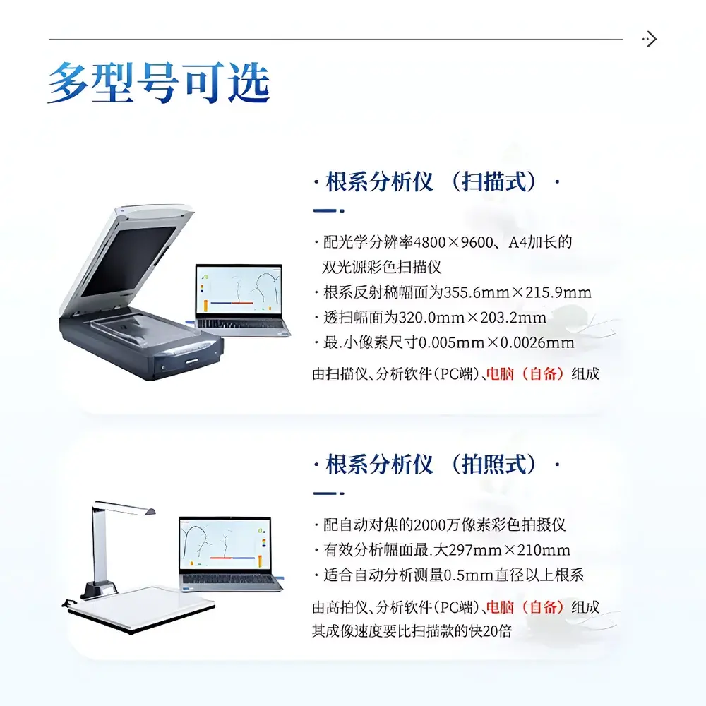
| Optical Resolution | 4800 × 9600 dpi (A4+ extended scanning area) |
| Reflective Scan Area | 355.6 mm × 215.9 mm |
| Transmissive Scan Area | 320.0 mm × 203.2 mm |
| Minimum Pixel Size | 0.005 mm × 0.0026 mm |
| Interface | USB 2.0 |
| Color Depth | 48-bit |
| Supported Image Formats | TIFF, JPEG |
| Software Licensing | Hardware-encrypted USB dongle |
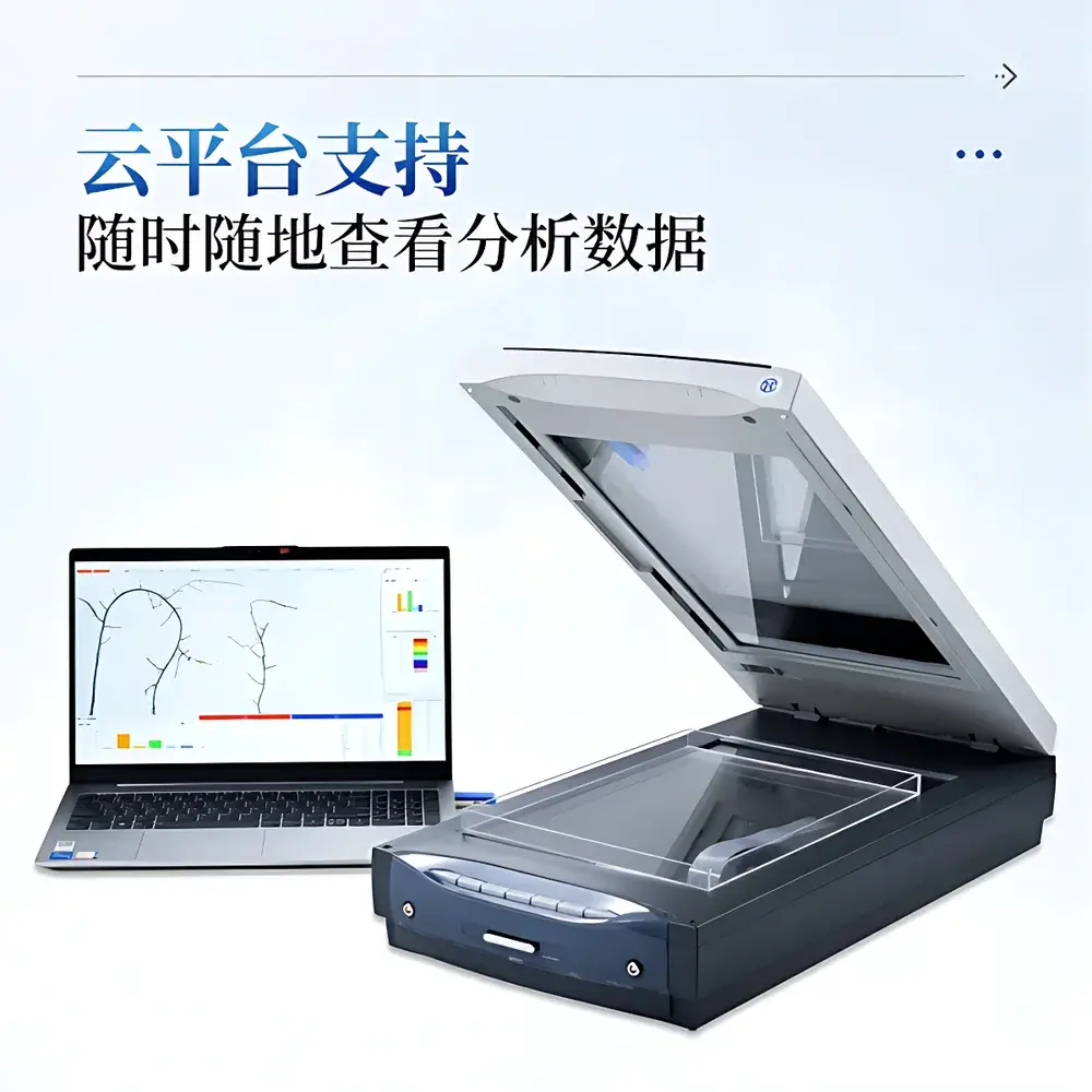
| Cloud Data Sync | Integrated web-based platform with encrypted HTTPS API |
| OS Compatibility | Windows 10 Professional / Enterprise (64-bit) |
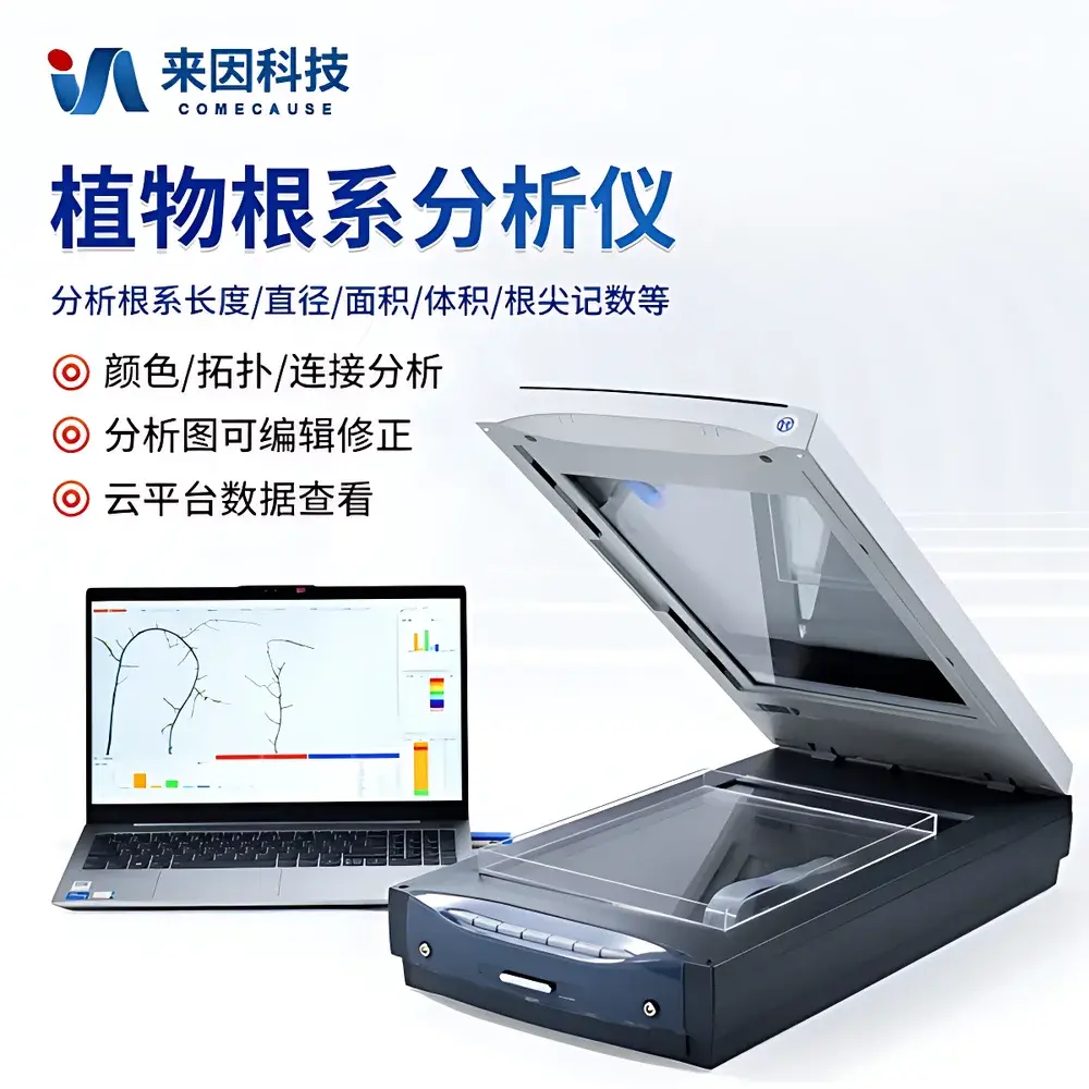
Overview
The COMECAUSE IN-GX02 Advanced Root System Analyzer is a dedicated benchtop imaging and morphometric analysis platform engineered for quantitative root phenotyping in controlled-environment plant science. It operates on the principle of high-fidelity optical digitization followed by deterministic image segmentation and skeleton-based topological reconstruction — not statistical sampling or machine-learning inference. The system captures root architecture via dual-illumination, A4+ extended-format reflective/transmissive scanning, eliminating shadow artifacts and ensuring uniform contrast across heterogeneous root samples (e.g., fine lateral roots, thick taproots, root nodules, or mycorrhizal hyphae). Its core analytical engine implements non-stochastic, pixel-accurate algorithms compliant with ISO 11277 (soil and root imaging standards) and aligned with FAO/CGIAR root trait ontology frameworks. Designed for reproducible longitudinal monitoring, it supports both ex situ washed-root analysis and in situ rhizobox-grown specimens, enabling rigorous comparison of root architectural plasticity under abiotic stress, nutrient gradients, or symbiotic treatments.
Key Features
- Dual-source CCFL illumination system with built-in calibration zones ensures consistent radiometric response across full scan area — critical for cross-experiment comparability.
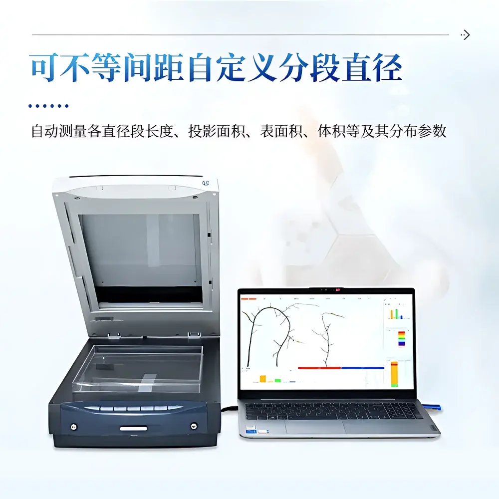
- A4+ extended scanning bed (355.6 × 215.9 mm reflective; 320.0 × 203.2 mm transmissive) accommodates large root systems without tiling or stitching artifacts.
- Hardware-encrypted software license (USB dongle) enforces traceable user access and audit-ready session logging per GLP requirements.
- Sub-5 µm effective pixel resolution (0.005 mm × 0.0026 mm) enables reliable detection of root hairs ≥15 µm diameter and precise delineation of cortical cell layers in cleared samples.
- Full morphometric suite: total length, diameter distribution (user-defined non-uniform bins), surface area, volume, tip count, branching order, intersection density, and fractal dimension (box-counting method).
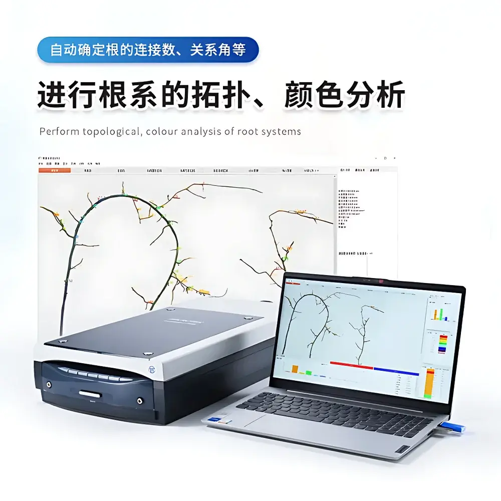
- Topology-aware analysis: automatic root hierarchy assignment, connection matrix generation, angle quantification (gravitropic & azimuthal), and isolateable sub-network extraction (e.g., primary root only, or nth-order laterals).
- Color-based segmentation module supports differential analysis of nitrogen-fixing nodules, pathogen-infected zones, or senescing tissues using calibrated RGB/HSL thresholds.
- Batch processing mode with configurable QC flags allows automated re-analysis of flagged images without manual intervention — validated for >500-sample datasets.
Sample Compatibility & Compliance
The IN-GX02 accepts intact root systems from hydroponic, agar-plate, rhizobox, or soil-wash protocols. Transparent acrylic root trays (included, n=3) are optically matched to minimize refraction distortion during transmissive scanning. The system meets ISO/IEC 17025 preconditions for measurement uncertainty estimation in morphometric imaging, and its software architecture supports 21 CFR Part 11-compliant electronic signatures when deployed with enterprise authentication (LDAP/Active Directory integration optional). All output metrics — including fractal dimension (Df) and root-shoot allocation ratios — are traceable to NIST-traceable calibration targets scanned alongside biological samples.
Software & Data Management
Analysis is performed via COMECAUSE RootVision™ v4.x, a native Windows application with modular workflow scripting. Raw scans are stored as lossless TIFFs with embedded EXIF metadata (timestamp, scanner ID, illumination mode). Processed results export to structured CSV/Excel with column headers conforming to MIAPPE 1.1 metadata standards. Cloud synchronization uses TLS 1.3-encrypted RESTful API endpoints; all data-at-rest is AES-256 encrypted. Audit trails record operator ID, timestamp, parameter set version, and image modification history — fully compatible with GxP-aligned lab information management systems (LIMS).
Applications
- Quantitative genetic mapping of root architecture QTLs under drought or low-phosphorus conditions.
- Phenotypic validation of CRISPR-edited root gravitropism mutants (e.g., arf7/arf19, pgm1).
- Time-series analysis of nodulation kinetics in Medicago truncatula–sinorhizobium symbiosis.
- Root system efficiency modeling for crop ideotype development (e.g., deep rooting index, lateral density gradient).
- Standardized root trait reporting for international germplasm screening initiatives (e.g., IWG-Root, EPPN2020).
FAQ
Does the IN-GX02 require proprietary consumables or subscription-based software updates?
No. All analysis modules are included in the perpetual license. Firmware and software updates are provided free-of-charge for hardware-supported versions.
Can the system analyze roots embedded in transparent gels or agar plates?
Yes — transmissive scanning mode resolves roots in 0.8–1.2% agarose or phytagel matrices without dehydration or staining.
Is raw image data export supported for third-party algorithm development?
Yes. Full-resolution TIFFs with embedded spatial calibration metadata are directly accessible via file system or API.
What validation documentation is supplied for regulatory submissions?
Includes IQ/OQ protocol templates, measurement uncertainty reports per GUM (JCGM 100:2008), and software verification test suites.
How is geometric distortion corrected across the extended scan field?
Through factory-calibrated lens distortion maps applied during image registration — verified using NIST SRM 2036 grid targets.


