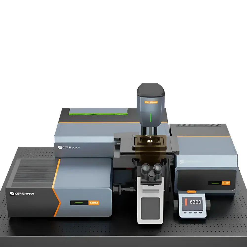
CSR Biotech Cell Xtreme Dual-Mode Live-Cell Super-Resolution Microscope
| Brand | CSR Biotech |
|---|---|
| Origin | Guangdong, China |
| Model | Cell Xtreme |
| Technology | Structured Illumination Microscopy (SIM) & Multi-Illumination Optical Diffraction Tomography (MI-ODT) |
| Resolution | XY: 60 nm, Z: 200 nm |
| Field of View | MI-ODT: up to 138 × 138 µm @100× |
| MI-SIM | up to 150 × 150 µm @100× |
| Imaging Speed | up to 25 fps @16-bit, 4608 × 4608 pixels |
| Eyepiece Options | Olympus IX85 / IX83 / Nikon Ti2-E |
| Objective | Apo 100×/1.5 Oil |
| Light Source | Multi-color coupled laser illumination system |
Overview
The CSR Biotech Cell Xtreme is a dual-mode, live-cell super-resolution microscope engineered for quantitative, long-term, and label-free or fluorescence-enhanced structural interrogation of dynamic biological systems. It integrates two complementary, co-aligned optical modalities—Multi-Illumination Structured Illumination Microscopy (MI-SIM) and Multi-Illumination Optical Diffraction Tomography (MI-ODT)—within a single, unified platform. Unlike conventional super-resolution instruments constrained to either fluorescent labeling or limited label-free contrast, the Cell Xtreme enables simultaneous acquisition of refractive index (RI) distribution maps via MI-ODT and molecular-specific spatial localization via MI-SIM. This dual-path architecture leverages orthogonal physical contrast mechanisms: MI-ODT reconstructs 3D RI tomograms from interferometric intensity measurements under multi-angle, multi-frequency illumination, while MI-SIM achieves ~2× resolution enhancement over diffraction-limited widefield imaging through patterned excitation and computational reconstruction. Both modalities operate under physiological conditions—including temperature-, CO₂-, and humidity-controlled environments—and are optimized for extended time-lapse acquisition with sub-second temporal resolution.
Key Features
- Dual-modal optical architecture: Co-registered MI-SIM and MI-ODT paths share a common sample plane and coordinate frame, enabling pixel-perfect fusion without post-acquisition alignment artifacts.
- Real-time dual-modality control: Integrated hardware synchronization allows concurrent or interleaved acquisition at up to 25 fps (MI-SIM) and 15–20 Hz (MI-ODT), with AI-assisted real-time segmentation and decision-triggered modality switching.
- High-stability illumination升降 module: Custom-designed laser illumination arm featuring closed-loop thermal regulation (±0.1 °C), dual-rail precision positioning (<1 µm repeatability, <100 µrad tilt error), and 500-position/sec programmable beam steering (<200 µs switching latency).
- Adaptive imaging orchestration: Supports multi-point autofocus, multi-location tracking, event-triggered acquisition, non-uniform z-stack scheduling, and multi-stage variable-frequency protocols—all configurable via graphical workflow editor.
- Physiological sample environment: Fully enclosed chamber with thermally conductive borosilicate coverslip and external objective-mounted baffles ensures stable long-term culture during high-NA imaging.
- Modular optical compatibility: Interchangeable objectives (100×/1.5 oil, 60×/1.4 oil, 60×/1.35 silicone, 60×/1.2 water) and expandable laser combiner support multi-channel spectral flexibility and future upgrade paths.
Sample Compatibility & Compliance
The Cell Xtreme accommodates adherent and suspension mammalian cells, primary neurons, organoids, and thin tissue explants (≤100 µm thickness). Its non-invasive MI-ODT modality eliminates phototoxicity and photobleaching concerns, making it suitable for longitudinal studies requiring >24-hour observation windows. The MI-SIM path supports standard fluorophores (e.g., Alexa Fluor 488, mCherry, SiR-tubulin) and is compatible with genetically encoded tags (e.g., GFP, mNeonGreen). System design complies with ISO 13485 quality management principles for laboratory instrumentation. Data provenance, user access logs, and audit trails are maintained in accordance with GLP and GMP-aligned workflows. Optional FDA 21 CFR Part 11-compliant electronic signature and e-record modules are available for regulated preclinical research environments.
Software & Data Management
The Cell Xtreme operates via OIA (Online Image Analysis) software—a modular, Python-based platform supporting real-time visualization, on-the-fly AI segmentation (e.g., nuclear envelope, mitochondria, lipid droplets, plasma membrane), and adaptive acquisition logic. Raw data is stored in standardized HDF5 format with embedded metadata (acquisition parameters, calibration references, environmental logs). The software includes built-in tools for SIM/ODT reconstruction, deconvolution, drift correction, and multi-modal registration. Export options include TIFF stacks, N5/Zarr volumes, and OMERO-compatible annotations. All processing pipelines are containerized (Docker) and reproducible across local workstations or HPC clusters. Remote monitoring and collaborative analysis are supported via secure WebSocket API and RESTful endpoints.
Applications
- Dynamic organelle interactions: Simultaneous tracking of mitochondrial fission/fusion, lysosome–autophagosome docking, and ER–mitochondria contact sites using MI-ODT-defined morphology + MI-SIM-labeled markers.
- Label-free phenotypic screening: Quantitative morphokinetic profiling of drug-treated cells—e.g., changes in nuclear condensation, cytoplasmic density gradients, or lipid droplet dynamics—without transfection or staining.
- Structural virology: High-speed MI-ODT captures virus entry and trafficking; MI-SIM resolves capsid protein organization and host factor recruitment at sub-100-nm scale.
- Neuronal development: Time-resolved mapping of growth cone advance, microtubule polymerization, and synaptic vesicle clustering in primary cultures under physiological buffer conditions.
- 3D cell migration in hydrogels: Combined MI-ODT volumetric RI reconstruction and MI-SIM actin/matrix labeling enable correlative analysis of mechanical deformation and cytoskeletal remodeling.
FAQ
What distinguishes MI-ODT from conventional ODT implementations?
MI-ODT introduces multi-illumination angular diversity, high-fidelity wavefront calibration via self-referencing targets, and real-time power-spectrum-based k-vector estimation—enabling robust tomographic reconstruction even under low-contrast, heterogeneous, or moving samples.
Can MI-SIM and MI-ODT be acquired simultaneously?
Yes—the system employs synchronized dual-camera readout and shared stage/illumination control, allowing true concurrent acquisition with hardware-level timestamp alignment.
Is the Cell Xtreme compatible with existing incubation chambers?
It supports integration with commercial stage-top incubators; however, its native sealed chamber provides superior thermal stability and optical access for high-NA objectives.
How is data integrity ensured during long-term acquisitions?
All acquisitions log environmental telemetry (temperature, CO₂, focus drift), illumination power, and hardware status in real time. Data files embed SHA-256 checksums and versioned reconstruction parameters.
Does the system support third-party analysis plugins?
Yes—OIA exposes a documented Python SDK and plugin interface compliant with scikit-image, napari, and DeepImageJ standards.


