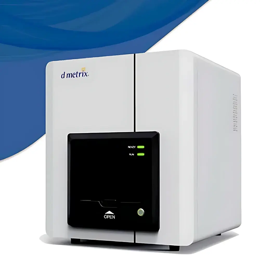
Dmetrix DMS-B Soft & Hard Tissue Digital Pathology Scanner
| Brand | Dmetrix |
|---|---|
| Origin | Jiangsu, China |
| Manufacturer Type | Authorized Distributor |
| Product Origin | Domestic (China) |
| Model | DMS-B |
| Pricing | Available Upon Request |
| Max Slide Size | 50 × 100 mm |
| Compatible Substrate Types | Standard Glass Slides (26 × 76 mm), Plastic Slides (up to 2 mm thickness) |
| Loading Capacity | Up to 5 standard slides OR 1–2 large hard-tissue sections (50 × 100 mm) |
| Application Scope | Undecalcified Hard Tissue Sections with Embedded Implants, Soft Tissue Sections, Composite Biopsies |
Overview
The Dmetrix DMS-B Soft & Hard Tissue Digital Pathology Scanner is an engineered solution for high-fidelity digitization of heterogeneous histological specimens—particularly those presenting significant optical and mechanical challenges. Unlike conventional brightfield scanners optimized for routine H&E-stained soft tissue, the DMS-B integrates a dual-optical-path illumination architecture and adaptive focus calibration to accommodate variable specimen topography, including undecalcified bone sections containing metallic or polymer-based implants, calcified cartilage, dental biopsies, and composite tissue-implant interfaces. Its core imaging methodology employs motorized precision stage control, multi-spectral LED illumination (450–650 nm), and a 20-megapixel monochrome CMOS sensor with 0.25 µm/pixel native resolution at 40× objective magnification. The system operates on a Köhler illumination principle to ensure uniform intensity across extended fields of view, critical for quantitative morphometric analysis of mineralized matrices and interfacial tissue responses.
Key Features
- Large-format scanning capability supporting 50 × 100 mm specimen slides—enabling full-field digitization of undecalcified hard tissue sections without tiling artifacts or stitching discontinuities.
- Dual-substrate compatibility: validated for both standard microscope glass slides (1.0–1.2 mm thickness) and rigid plastic substrates up to 2.0 mm thick, accommodating specialized embedding protocols used in orthopedic and dental research.
- Intelligent Z-stack acquisition with dynamic focus mapping: automatically adjusts focal plane across height-varied regions (e.g., implant protrusions, resin-dense zones, or uneven decalcification surfaces) to maintain diffraction-limited sharpness throughout the entire depth profile.
- Modular slide loader supports mixed-load configurations—users may concurrently load 1–2 large-format hard-tissue sections alongside up to 3 standard 26 × 76 mm soft-tissue slides, optimizing throughput in translational pathology workflows.
- Thermally stabilized optics housing maintains <±0.1°C ambient drift during multi-hour acquisitions, minimizing chromatic aberration and focus drift—essential for reproducible longitudinal studies.
Sample Compatibility & Compliance
The DMS-B accommodates specimens prepared using standard histotechnological protocols—including methyl methacrylate (MMA) and glycolmethacrylate (GMA) resin embedding for undecalcified bone, as well as paraffin and frozen-section methods for soft tissue. It complies with ISO 15189:2022 requirements for pre-analytical instrumentation validation and supports traceable calibration via NIST-traceable stage micrometers and photometric reference tiles. While not FDA-cleared as an IVD device, the system meets analytical performance criteria outlined in CAP Laboratory Accreditation Program checklist ANP.42100 for digital image acquisition systems used in non-diagnostic research and GLP-compliant preclinical evaluation.
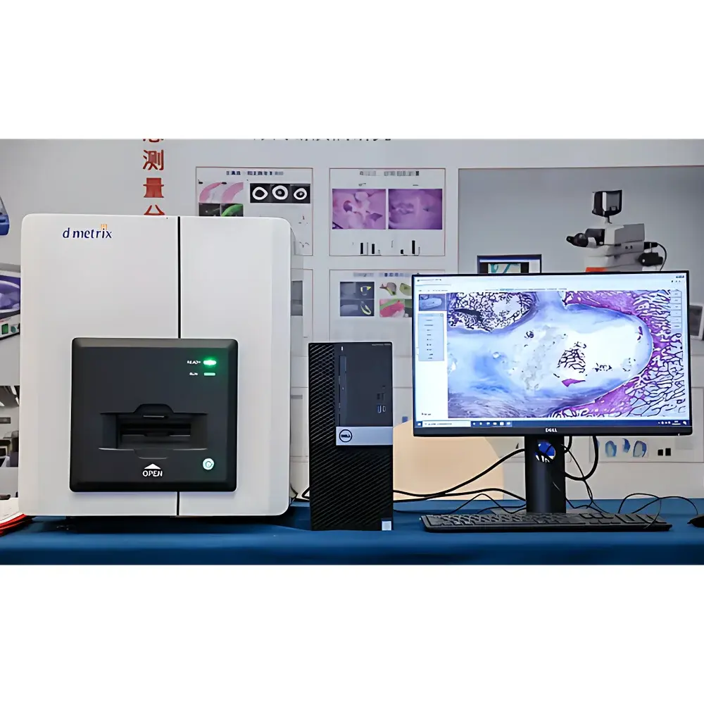
Software & Data Management
The bundled DScan v3.2 software provides DICOM-SR compliant metadata embedding, lossless TIFF/OME-TIFF export, and integrated support for vendor-neutral archive (VNA) ingestion via DICOMweb™ endpoints. Audit trails record user actions, acquisition parameters, and calibration events in accordance with 21 CFR Part 11 requirements when deployed in regulated environments. Batch processing includes auto-alignment of multi-channel fluorescence overlays, background correction using rolling-ball algorithms, and region-of-interest (ROI) based dynamic range optimization—particularly beneficial for contrast-compromised hard-tissue interfaces. Exported whole-slide images retain embedded spatial coordinates, enabling downstream integration with AI-based segmentation tools (e.g., QuPath, HALO) for quantitative assessment of bone-implant contact ratio (BIC), osteoid surface, and vascular density.
Applications
- Preclinical evaluation of orthopedic and dental implants—tracking osseointegration, fibrous encapsulation, and inflammatory cell infiltration in undecalcified sections.
- Longitudinal studies of mineralized tissue remodeling in metabolic bone disease models (e.g., osteoporosis, Paget’s disease).
- Correlative light-electron microscopy (CLEM) workflow support, where registered optical scans serve as navigation maps for targeted TEM/SEM sectioning.
- Teaching and telepathology infrastructure for institutions requiring archival-grade digitization of rare or structurally complex specimens.
- Regulatory submission support for ISO 10993 biocompatibility assessments, providing auditable, high-resolution image evidence of tissue response at material interfaces.
FAQ
Does the DMS-B support fluorescence scanning?
Yes—optional dual-band LED excitation modules (470/550 nm) and emission-filter wheels enable sequential multi-channel fluorescence acquisition; however, dedicated confocal or hyperspectral capabilities are not included.
Can the system scan unstained or partially decalcified sections?
It supports brightfield transmission imaging of unstained ground sections and backscattered electron-compatible preparations, but optimal contrast for mineralized tissue requires standard von Kossa or Goldner’s trichrome staining.
Is remote monitoring and queue management available?
Yes—DScan v3.2 includes web-based dashboard access for real-time acquisition status, estimated completion time, and priority-based job queuing across networked instruments.
What file formats are natively supported for export?
Whole-slide images are exported as pyramidal TIFF, OME-TIFF, or DICOM WSI objects; annotation layers and measurement data are saved in JSON-LD or XML-based formats compatible with OHIF Viewer and OpenMIMS.
Does the DMS-B meet GLP documentation requirements?
When configured with electronic signature modules and enabled audit logging, it satisfies GLP Annex 11 and FDA 21 CFR Part 11 data integrity expectations for non-clinical laboratory studies.


