Echo Revolve RVL-100-G Phase Contrast Microscope
| Brand | ECHO |
|---|---|
| Origin | USA |
| Model | RVL-100-G |
| Configuration | Upright/Inverted Convertible Microscope |
| Optical Modes | Brightfield, Darkfield, Phase Contrast, Fluorescence, Polarization |
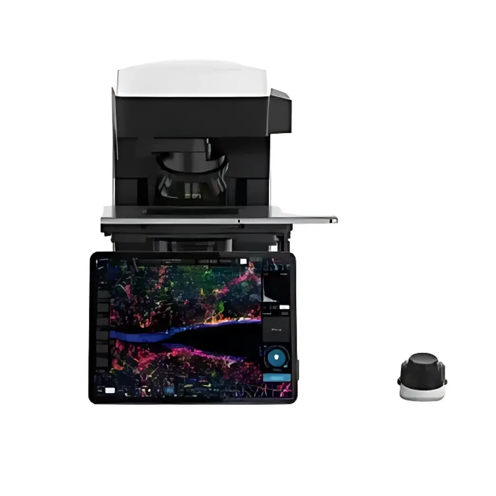
| Display | 12.9-inch Retina Touchscreen (iPad Pro-based) |
| Objective Compatibility | Olympus® 1.25x–100x achromat, fluorite, and apochromat objectives |
| Condenser | Long-working-distance & high-resolution condensers |
| Fluorescence Illumination | 5-channel high-intensity LED system (50,000 hr lifetime) |
| Cameras | Dual-sensor imaging — 12 MP color CMOS (BF), 5 MP monochrome sCMOS (FL) |
| Digital Image Processing | Real-time GPU-accelerated Digital Haze Reduction (DHR) |
Overview
The Echo Revolve RVL-100-G is a fully integrated upright/inverted convertible phase contrast microscope engineered for advanced life science research and routine cell biology applications. Unlike conventional modular systems, the Revolve employs a unified optical and control architecture that eliminates mechanical reconfiguration—users seamlessly transition between upright and inverted configurations via a single slide mechanism, preserving parfocality and optical alignment without recalibration. Its core optical design adheres to Köhler illumination principles across all transmitted-light modes (brightfield, darkfield, phase contrast, and polarization), while fluorescence imaging leverages epi-illumination with precisely aligned 5-channel LED excitation sources. The system’s phase contrast capability utilizes annular diaphragms and phase-shifted objectives (compatible with standard Olympus® phase objectives) to enhance contrast in unstained, transparent biological specimens—enabling real-time observation of live cells in culture dishes, multi-well plates, or glass slides without fixation or staining. Designed for compliance with ISO 10993 biocompatibility guidelines for optical components in contact with biological samples, the Revolve supports GLP-aligned workflows through audit-trail-capable software logging.
Key Features
- Upright/inverted convertible platform: One mechanical slide transforms optical path geometry—upright mode optimized for glass slide imaging; inverted mode configured for bottom-mounted culture vessels (e.g., Petri dishes, 6–96-well plates, T-flasks) with ≥2 mm working distance.
- Multi-modal illumination engine: Integrated LED-based brightfield, darkfield, phase contrast, polarized, and fluorescence illumination—all independently controllable via touchscreen interface with calibrated intensity ramping (0.1–100% stepless dimming).
- Dual-camera acquisition system: Simultaneous or sequential capture using a 12-megapixel color CMOS sensor (for brightfield documentation and color phase contrast) and a scientific-grade 5-megapixel monochrome sCMOS sensor (optimized for low-noise, high-dynamic-range fluorescence detection).
- Retina-display control interface: 12.9-inch iPad Pro serves as embedded controller—running proprietary macOS-derived imaging software with native support for touch gestures, pinch-to-zoom, swipe-driven mode switching, and on-device image annotation.
- Digital Haze Reduction (DHR): GPU-accelerated real-time deconvolution algorithm applied during acquisition to suppress out-of-focus blur and photon scatter—particularly effective for thick specimens (>20 µm) and widefield fluorescence imaging where axial resolution is inherently limited.
- All-in-one compact chassis: No external PC, power supply rack, or lamp housing required. Wi-Fi 5 (802.11ac) enables wireless synchronization between host optics and display; magnetic mount allows flexible tablet positioning without obstructing ergonomics.
Sample Compatibility & Compliance
The Revolve RVL-100-G accommodates diverse specimen formats without hardware modification: standard 1″ × 3″ glass slides (upright), 35–150 mm Petri dishes, 6–384-well microplates, and T-25 to T-225 flasks (inverted). Stage travel (X/Y: 76 × 52 mm) and Z-focus range (≥10 mm) support high-throughput screening and time-lapse imaging across heterogeneous sample geometries. All optical surfaces comply with ISO 8578 (microscope objective labeling) and ANSI Z80.10 (ophthalmic and biomedical optical safety). The LED fluorescence module meets IEC 62471 photobiological safety Class 1 requirements. Software complies with FDA 21 CFR Part 11 for electronic records and signatures when operated in audit-mode configuration—supporting user-defined roles, password-protected method locking, and immutable acquisition logs.
Software & Data Management
The Revolve Control Suite runs natively on iPadOS and interfaces directly with the microscope’s embedded ARM-based imaging controller. It supports DICOM-compliant export (DICOM-SR for structured reporting), TIFF/OME-TIFF metadata embedding (including objective ID, exposure time, gain, LED channel activation), and direct cloud upload to secure HIPAA-compliant storage endpoints (AWS S3, Microsoft Azure Blob). Batch processing pipelines include flat-field correction, chromatic aberration compensation, and DHR parameter tuning per channel. Exported datasets retain full EXIF-like metadata for traceability in regulatory submissions (e.g., USP <1043>, ISO/IEC 17025 calibration documentation).
Applications
- Live-cell phase contrast imaging of adherent and suspension cultures over extended durations (e.g., wound-healing assays, mitosis tracking).
- Multi-parameter phenotypic screening in 96-well plates using combined phase contrast + fluorescence (e.g., GFP-tagged organelles + nuclear DAPI counterstain).
- Quality control of primary cell isolates and stem cell differentiation protocols under GMP-aligned environments.
- Teaching laboratories requiring intuitive operation for undergraduate microscopy instruction—eliminating complex alignment procedures typical of traditional compound scopes.
- Preclinical imaging of explanted tissues (e.g., corneal epithelium, intestinal organoids) where rapid modality switching between phase and fluorescence is essential.
FAQ
Does the Revolve RVL-100-G support third-party objective lenses?
Yes—it accepts standard RMS-threaded (Royal Microscopical Society) objectives with 45 mm parfocal distance, including Olympus® UPlanSApo, Nikon CFI Plan Apo, and Zeiss EC Epiplan series. Mechanical stop rings prevent over-insertion.
Is the DHR algorithm compatible with post-acquisition analysis?
No—DHR operates exclusively in real-time acquisition mode. Raw unprocessed frames are not accessible; however, the system saves both DHR-processed and intermediate dehazed buffers for comparative validation.
Can the system be integrated into existing LIMS or ELN platforms?
Yes—via RESTful API endpoints documented in the Revolve Developer SDK, supporting automated job queuing, metadata ingestion, and instrument status polling.
What is the maximum supported magnification for phase contrast imaging?
With the included 100× oil-immersion apochromat objective and 1.35 NA condenser, theoretical resolution is ≤220 nm (Abbe limit); practical phase contrast contrast transfer extends to ~0.5 µm features in live cytoplasmic structures.
How is focus stability maintained during long-term time-lapse experiments?
A motorized Z-drive with closed-loop piezoelectric feedback maintains focus drift within ±0.2 µm over 24 hours at ambient temperature (22 ± 2°C), validated per ASTM E2925-19 Annex A2.


