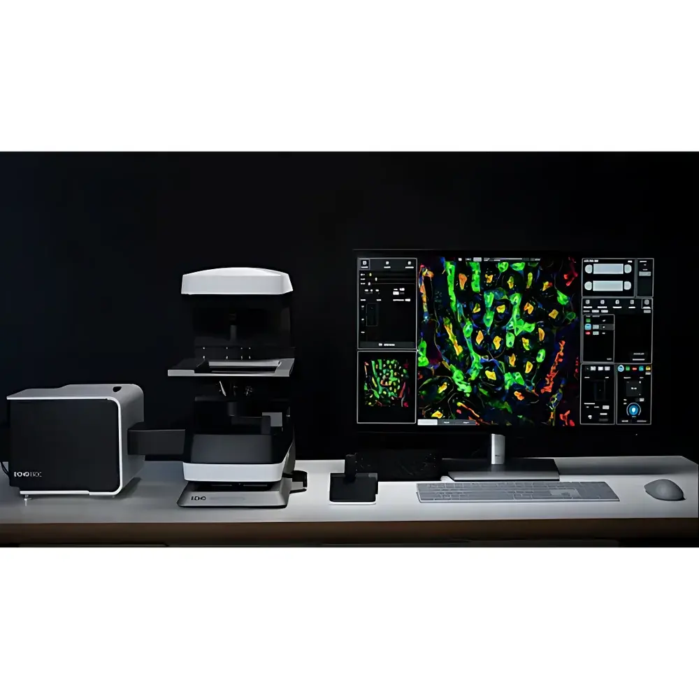
ECHO SDC Confocal Spinning Disk Microscope
| Brand | ECHO |
|---|---|
| Origin | USA |
| Manufacturer Type | Authorized Distributor |
| Product Category | Imported Instrument |
| Model | SDC |
| Configuration | Upright/Inverted Integrated Platform |
| Imaging Modality | Spinning Disk Confocal, Widefield Fluorescence, Super-Resolution (SIM-based) |
Overview
The ECHO SDC Confocal Spinning Disk Microscope is an integrated upright/inverted platform engineered for high-speed, low-phototoxicity fluorescence imaging of live biological specimens. Based on the optical principle of spinning disk confocal microscopy—where a rotating Nipkow disk with thousands of precisely aligned microlens-pinhole pairs enables parallel illumination and detection—the system achieves true optical sectioning without point-scanning latency. Unlike laser scanning confocal microscopes (LSCM), which acquire images serially pixel-by-pixel, the SDC captures full-frame confocal data at up to 200 frames per second (fps), enabling real-time visualization of dynamic subcellular processes including intracellular Ca2+ transients, vesicle trafficking, mitochondrial dynamics, and hemodynamic flow in thick tissue preparations. Its optical architecture incorporates conjugate pinhole filtering at the image plane to reject out-of-focus light, delivering enhanced axial resolution (~0.7 µm) and improved signal-to-noise ratio compared to widefield fluorescence, while maintaining compatibility with standard fluorophores and genetically encoded indicators (e.g., GFP, mCherry, tdTomato).
Key Features
- Integrated Dual-Mode Platform: Seamless switching between widefield fluorescence and spinning disk confocal modalities on a single, rigid mechanical base—eliminating re-alignment and optical path recalibration.
- High-Speed Full-Frame Acquisition: Capable of 200 fps confocal imaging at 120 nm effective resolution (structured illumination-enhanced mode), supporting time-lapse studies of rapid physiological events.
- Reduced Photodamage Architecture: Low-intensity, parallelized excitation minimizes cumulative photobleaching and phototoxic stress—validated for >6-hour continuous imaging of sensitive primary neurons and stem cell cultures.
- Compact, Cable-Managed Design: Optimized footprint (< 0.8 m²) with internalized optical routing and minimal external cabling—designed for shared core facilities and BSL-2 laboratory environments.
- Intuitive Workflow Software: Unified acquisition interface with one-click modality selection, automated multi-channel registration, Z-stack tiling, and real-time deconvolution preview.
Sample Compatibility & Compliance
The ECHO SDC supports diverse live and fixed specimen formats: adherent mammalian cells (glass-bottom dishes, chamber slides), 3D organoids (up to 200 µm thickness), zebrafish embryos (mounted in low-melt agarose), and ex vivo brain slices. It complies with ISO 13485–certified manufacturing protocols for medical device components and meets key regulatory prerequisites for GLP-compliant imaging workflows—including audit-trail-enabled metadata logging (acquisition parameters, timestamps, user IDs) and electronic signature support aligned with FDA 21 CFR Part 11 requirements. All optical components are certified to RoHS and REACH standards; environmental controls meet ASTM E1545 specifications for incubator performance validation.
Software & Data Management
Acquisition and analysis are managed via ECHO Vision Suite v4.x—a modular, Windows-based platform compliant with MIAME and OME-TIFF open-data standards. Core modules include: (1) AutoFocus Pro with adaptive contrast-based Z-lock for long-term timelapse stability; (2) MultiChannel Sync, enabling hardware-triggered synchronization across four independent LED/Laser sources (405/488/561/640 nm); (3) Z-Stack Composer, supporting automatic drift correction, intensity normalization, and maximum-intensity projection export; and (4) Quantify+ Plugin, providing batch ROI analysis, colocalization metrics (Pearson’s r, Mander’s coefficients), and kinetic curve fitting (exponential decay, Hill equation). Raw data is stored in vendor-neutral OME-Zarr format with embedded JSON metadata for FAIR data principles adherence.
Applications
- Live-cell calcium signaling kinetics in cardiomyocytes and neuronal networks
- Real-time tracking of endosomal trafficking and lysosomal pH dynamics
- 3D reconstruction of vascular networks in tumor spheroids
- Time-resolved imaging of mitotic spindle assembly and chromosome segregation
- Super-resolution structural mapping of nuclear pore complexes and cytoskeletal remodeling
- Long-term lineage tracing in embryonic stem cell differentiation assays
FAQ
What is the maximum supported Z-stack depth and acquisition speed?
Up to 500 optical sections at 200 fps in confocal mode; speed scales inversely with section count and resolution binning.
Does the system support objective correction collars for temperature-induced refractive index shifts?
Yes—motorized correction collars are available for all 40× and 60× water-immersion objectives, calibrated for 20–40 °C operation.
Can third-party cameras be integrated via GenICam or DCAM protocols?
Native support for Hamamatsu ORCA-Fusion BT and Photometrics Prime BSI Express cameras; GenICam-compliant models require firmware-level validation by ECHO Applications Engineering.
Is humidity control validated per ISO 8573-1 Class 4 specifications?
Yes—the integrated incubation module maintains 85–95% RH ±2% over 72 h, with gravimetric calibration traceable to NIST SRM 2689a.
How is photobleaching quantified during acquisition?
ECHO Vision Suite logs per-channel photon flux (photons/pixel/frame) and computes normalized bleaching rates using exponential decay modeling against baseline intensity.


