FluidicLab Flow Cell for Laminar Flow Perfusion Systems
| Brand | FluidicLab |
|---|---|
| Origin | Shanghai, China |
| Manufacturer Type | Direct Manufacturer |
| Region of Origin | Domestic (China) |
| Model | Flow Cell |
| Price | USD 1,350 (approx.) |
| Instrument Type | Manual |
| Dimensions (L×W×H) | 60 × 65 × 15 mm |
Overview
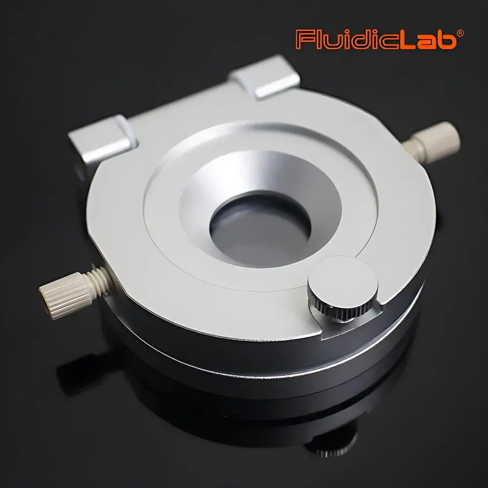
The FluidicLab Flow Cell is a precision-engineered laminar flow perfusion chamber designed for integration into life science instrumentation platforms requiring controlled, low-shear liquid delivery and real-time optical interrogation. Based on the principle of hydrodynamic focusing and Poiseuille flow, this device enables stable, predictable laminar transport of biological samples—such as suspended cells, nucleic acids, or protein complexes—across an optically accessible path. Its sandwich architecture ensures minimal dead volume, rapid solution exchange (<1 s for full chamber turnover under standard flow rates), and compatibility with high-resolution microscopy modalities including epifluorescence, confocal, and TIRF. Unlike conventional cuvettes or static wells, the Flow Cell supports dynamic concentration gradients, sequential reagent switching, and continuous sample refresh—critical for electrophysiology assays, single-cell imaging, hybridization kinetics, and microarray-based functional screening.
Key Features
- Sandwich-style modular construction: removable #1.5 coverglass (Ø40 mm, 0.17 mm thick), customizable silicone gasket layer (standard thicknesses: 0.1 mm or 0.2 mm; geometry options include F2414 and 47815-F; custom profiles available upon request), and top-flow glass plate with integrated microfluidic inlet/outlet channels.
- Optical compatibility: fully transparent quartz-grade glass surfaces with <0.1 µm surface roughness; supports widefield, confocal, and super-resolution imaging across visible to near-UV spectra (200–800 nm).
- Microscope integration: designed for direct mounting on standard inverted microscope stages; accommodates 22 mm diameter field-of-view without mechanical interference; includes PEEK 10-32 threaded fittings (6 pcs) and 3 m PTFE capillary tubing (OD 1.6 mm) for leak-free fluidic interfacing.
- Reusable and sterilizable: silicone gaskets withstand autoclaving (121°C, 20 min, 15 psi); coverglasses are single-use but cost-effective (20 included per kit); gasket replacement is tool-free and takes <30 seconds.
- Low-volume operation: internal channel volume ranges from 1.2 µL (0.1 mm gasket) to 2.4 µL (0.2 mm gasket), minimizing reagent consumption while maintaining laminar Reynolds numbers <100 under typical flow rates (1–100 µL/min).
Sample Compatibility & Compliance
The Flow Cell is validated for use with aqueous buffers (PBS, HEPES, Tyrode’s), cell culture media (DMEM, RPMI), glycerol-based mounting media, and low-viscosity organic solvents (e.g., ethanol, isopropanol) up to 30% v/v. It is compatible with mammalian cells (adherent and suspension), bacteria, exosomes, DNA/RNA oligos, antibodies, and peptide arrays. All wetted materials—silicone elastomer (medical-grade PDMS alternative), borosilicate glass, and PEEK—meet USP Class VI biocompatibility standards and ISO 10993-5 cytotoxicity requirements. The device supports GLP-compliant experimental workflows when paired with traceable calibration protocols and audit-ready logbooks; while not FDA 21 CFR Part 11 certified as a standalone unit, it integrates seamlessly with compliant data acquisition systems supporting electronic signatures and secure audit trails.
Software & Data Management
As a passive hardware component, the Flow Cell requires no embedded firmware or proprietary drivers. It operates transparently with third-party control platforms—including MATLAB-based pump scripting, LabVIEW DAQ modules, Python-controlled syringe pumps (e.g., Harvard Apparatus PHD Ultra), and commercial electrophysiology suites (Axon pCLAMP, HEKA PatchMaster). Flow rate calibration is performed using gravimetric or fluorescent tracer methods per ASTM D3595-20. Image metadata (time stamps, flow conditions, stage coordinates) can be synchronized via TTL triggers or software API hooks. Raw image stacks acquired during perfusion are natively compatible with Fiji/ImageJ, Imaris, and commercial analysis pipelines for kymograph generation, intensity quantification, and co-localization mapping.
Applications
- Nucleic Acid Hybridization Sequencing: Enables kinetic monitoring of probe-target binding in real time; supports whole-genome association studies (GWAS) by maintaining uniform flow over immobilized oligonucleotide arrays.
- Gene Expression Profiling: Facilitates quantitative RNA detection via fluorescent in situ hybridization (FISH) under continuous buffer exchange, reducing background and improving signal-to-noise ratio in single-cell transcriptomics.
- Peptide & Antibody Microarrays: Allows high-throughput screening of protein–protein interactions (e.g., Pro-Pro binding affinities) with programmable multi-step washing and elution cycles.
- Transfection & Compound Screening: Delivers plasmid DNA, siRNA, or small-molecule libraries to localized cellular microenvironments while preserving viability—ideal for CRISPR perturbation assays and phenotypic drug discovery.
- Electrophysiological Perfusion: Provides rapid solution switching (<500 ms) for voltage-clamp or patch-clamp experiments requiring precise agonist/antagonist application without mechanical disturbance.
FAQ
Is the Flow Cell compatible with high-pressure microfluidic systems?
No—it is rated for pressures up to 100 kPa (14.5 psi); for higher-pressure applications, external pressure regulators or flow restrictors are recommended.
Can I modify the gasket geometry for non-standard channel widths?
Yes—custom gasket designs (including tapered, bifurcated, or multi-inlet configurations) are available through FluidicLab’s engineering support team with lead times of 2–3 weeks.
Does the device support temperature control?
The base Flow Cell is unheated; however, it can be mounted onto commercially available microscope-stage incubators (e.g., Tokai Hit INU series) or integrated with custom thermal plates using its standardized footprint.
What surface treatments are possible on the coverglass?
Standard plasma cleaning, silanization (e.g., APTES), PEGylation, fibronectin/laminin coating, and UV-ozone activation are all compatible with the supplied #1.5 coverglasses.
How often should the silicone gasket be replaced?
Under routine sterile use with aqueous buffers, gaskets maintain dimensional stability for ≥50 autoclave cycles; visual inspection for cracking or compression set is advised after 30 cycles.
Related Products


