Formulatrix MUVIS® Manual UV Fluorescence Imaging System
| Brand | Formulatrix |
|---|---|
| Origin | USA |
| Manufacturer Type | Original Equipment Manufacturer (OEM) |
| Import Status | Imported |
| Model | MUVIS® |
| Field of View (mm) | 3.7 × 3.0 |
| Pixel Size (µm) | 1.1 |
| Optical Resolution (µm) | 2 |
Overview
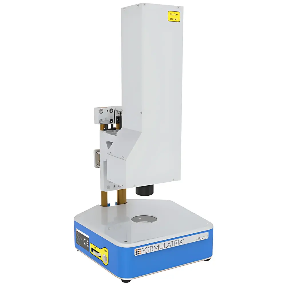
The Formulatrix MUVIS® Manual UV Fluorescence Imaging System is a compact, benchtop instrument engineered for rapid, non-destructive identification and documentation of protein crystals in crystallization screens. It operates on dual-modal optical detection: visible-light transmittance imaging for general morphology assessment and 280 nm ultraviolet (UV) fluorescence excitation to selectively visualize intrinsic tryptophan and tyrosine fluorescence—key spectral signatures of native protein structure. Unlike automated high-throughput platforms, the MUVIS® system adopts a manual, operator-guided workflow optimized for early-stage crystal screening in academic labs, core facilities, and contract research organizations where flexibility, footprint efficiency, and cost-conscious instrumentation are critical. Its design adheres to SBS (Society for Biomolecular Sciences) microplate standards and supports both standard polymer microplates and lipidic cubic phase (LCP) glass plates—enabling seamless integration into existing crystallization pipelines without protocol revalidation.
Key Features
- Compact form factor: Occupies only a 28 cm × 28 cm footprint and weighs under 5 kg—ideal for shared lab spaces, glove boxes, or mobile deployment across multiple crystallization rooms.
- Dual-mode optical imaging: Simultaneous support for white-light transmitted imaging (for plate layout, precipitant clarity, and crystal birefringence assessment) and 280 nm UV excitation with narrowband emission filtering (optimized for aromatic amino acid fluorescence at ~340–360 nm).
- High spatial fidelity: 1.1 µm pixel size and 2 µm optical resolution enable clear discrimination of sub-10 µm crystal features—including needle clusters, microcrystals, and twinned growth forms—without oil immersion or mechanical scanning.
- Manual precision control: Focus, exposure time, and LED intensity are manually adjustable via intuitive front-panel dials, eliminating software dependency during initial screening and enabling real-time parameter optimization per well.
- SBS- and LCP-compatible stage: Accepts 96-well, 384-well, and custom-format microplates; accommodates both standard polystyrene plates and fragile LCP glass sandwich plates up to 1.2 mm thickness.
Sample Compatibility & Compliance
The MUVIS® system is validated for use with aqueous and lipidic crystallization matrices containing common precipitants (e.g., PEGs, ammonium sulfate, MPD), cryoprotectants (e.g., glycerol, ethylene glycol), and additives (e.g., ligands, detergents). It imposes no thermal or vacuum requirements, preserving sample integrity during repeated imaging. While not classified as a regulated medical device, its optical architecture complies with IEC 62471 (Photobiological Safety of Lamps and Lamp Systems) for UV-A exposure limits. Data acquisition workflows align with GLP principles through timestamped image metadata (EXIF-compliant), supporting traceability in academic publications and preclinical structural biology studies.
Software & Data Management
The MUVIS® operates in standalone mode without proprietary software installation. All images are saved in lossless TIFF format with embedded metadata (wavelength, exposure time, focus position, plate ID). Optional integration with Formulatrix’s CrystalScore™ analysis suite (v3.2+) enables batch annotation, crystal scoring based on fluorescence intensity uniformity and edge sharpness, and export to CSV or JSON for downstream statistical analysis in Python or R. Audit trails—including user ID, session timestamps, and parameter logs—are retained locally on connected USB storage devices, satisfying basic 21 CFR Part 11 readiness when paired with institutional identity management systems.
Applications
- Rapid validation of protein crystal identity in sparse-matrix and grid screens—distinguishing protein crystals from salt or polymer artifacts via intrinsic fluorescence.
- Monitoring crystal growth kinetics across time-series experiments using consistent illumination and exposure settings.
- Pre-screening for synchrotron or cryo-EM beamtime allocation by prioritizing wells exhibiting strong, homogeneous UV signal and defined morphology.
- Teaching and training applications in structural biology courses—providing hands-on experience with fluorescence-based crystal detection without requiring advanced optics training.
- Supporting fragment-based drug discovery (FBDD) workflows by detecting ligand-induced stabilization of weakly diffracting crystals through enhanced fluorescence contrast.
FAQ
Does the MUVIS® require alignment or calibration before each use?
No. The fixed-focus optical path and factory-aligned UV LED module eliminate routine recalibration; only manual focus adjustment is needed per plate thickness.
Can the system detect nucleic acid crystals?
Limited utility: While DNA/RNA exhibit weak UV fluorescence at 280 nm, signal-to-noise ratio is significantly lower than for tryptophan-rich proteins; it is not recommended for primary nucleic acid crystal identification.
Is the UV source safe for repeated operator exposure?
Yes—the 280 nm LED emits <0.3 mW/cm² at the plate surface (measured at 25 cm working distance), well below IEC 62471 “Exempt” classification thresholds; no protective eyewear is required during normal operation.
How does MUVIS® compare to automated systems like Rock Imager or Rigaku CryoVista?
MUVIS® trades throughput and robotic plate handling for portability, immediate accessibility, and lower total cost of ownership—making it optimal for labs processing ≤50 plates/week or requiring on-demand imaging outside centralized facilities.
What file formats are supported for export and long-term archival?
Native TIFF with embedded EXIF tags; optional conversion to PNG or JPEG via third-party tools; metadata schema is publicly documented for institutional LIMS integration.
Related Products


