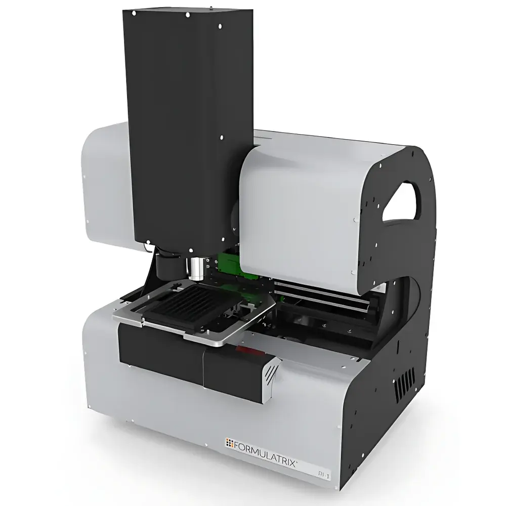
Formulatrix ROCK IMAGER 1® Protein Crystal Imager
| Brand | Formulatrix |
|---|---|
| Origin | USA |
| Manufacturer Type | Original Equipment Manufacturer (OEM) |
| Import Status | Imported |
| Model | RI1 |
| Field of View (mm) | 6.1 × 5.0 – 1.9 × 1.5 |
| Pixel Size (µm) | 1.8 – 0.56 |
| Optical Resolution (µm) | 5 – 1 |
Overview
The Formulatrix ROCK IMAGER 1® (RI1) is a compact, benchtop automated imaging system engineered specifically for high-fidelity documentation and longitudinal monitoring of protein crystallization experiments. It operates on a dual-modality optical architecture—combining high-resolution brightfield microscopy with UV-induced fluorescence excitation—to enable unambiguous differentiation between crystal growth, precipitate formation, phase separation, and amorphous aggregates. Unlike general-purpose microscopes or macro-imaging platforms, the RI1 is purpose-built for SBS-standard crystallization plates and integrates seamlessly into robotic crystallization workflows. Its modular optical train employs motorized zoom optics, precision-aligned LED illumination (470 nm and 365 nm), and a scientific-grade CMOS sensor with programmable exposure control—ensuring consistent, quantitative image acquisition across time-series experiments under controlled environmental conditions.
Key Features
- Motorized continuous zoom optics delivering variable field-of-view from 6.1 × 5.0 mm down to 1.9 × 1.5 mm—enabling both whole-plate survey imaging and high-magnification crystal-level inspection without manual lens changes.
- Scientific CMOS sensor with pixel sizes configurable between 1.8 µm and 0.56 µm, supporting optical resolution down to 1 µm at maximum magnification—sufficient to resolve lattice features in well-diffracting crystals.
- Dual-channel illumination: high-stability white LED (brightfield/phase-like contrast) and 365 nm UV LED for tryptophan-based intrinsic fluorescence detection—critical for distinguishing protein crystals from salt or polymer precipitates.
- Native compatibility with SBS-compliant plate formats including standard 96-well, Linbro, Nextal, and lipidic cubic phase (LCP) plates—accommodating both vapor diffusion and microbatch setups.
- Integrated environmental shielding with anti-condensation optics and optional humidity-controlled chamber interface—minimizing dew formation during long-term time-lapse imaging.
- Firmware-level trigger synchronization for external robotics (e.g., Tecan Freedom EVO, Hamilton STAR) enabling fully automated plate loading, focus calibration, and multi-position imaging sequences.
Sample Compatibility & Compliance
The RI1 accepts all commercially available crystallization plates adhering to the ANSI/SBS-1–2004 dimensional standard. It supports glass-bottom, polymer, and siliconized plastic plates—including specialized LCP sandwich plates with 10–20 µm thick membranes. No sample preparation or staining is required; imaging is performed in situ through plate lids or sealed films. The system complies with ISO 13485 design controls for medical device-related R&D instrumentation and meets electromagnetic compatibility (EMC) requirements per IEC 61326-1. All firmware and software components are architected to support audit trails and user access control—facilitating alignment with GLP and GMP documentation practices where crystallization data contributes to structural biology submissions.
Software & Data Management
The RI1 operates natively with ROCK MAKER™ v5.x, Formulatrix’s dedicated crystallization experiment management suite. This application provides automated focus mapping per well, dynamic exposure optimization, batch metadata tagging (e.g., condition ID, pH, precipitant concentration), and TIFF/PNG export with embedded EXIF-style acquisition parameters (exposure time, gain, LED intensity, zoom position). Exported images retain full spatial calibration metadata, enabling downstream integration with third-party crystal detection algorithms (e.g., CryoGrid, X-ray hit prediction pipelines). The software supports DICOM-SR structured reporting for institutional LIMS integration and includes built-in tools for time-lapse GIF generation, multi-well comparison overlays, and false-color intensity heatmaps aligned to crystallization screen grids. Data integrity is enforced via SHA-256 checksum logging and optional 21 CFR Part 11–compliant electronic signature modules.
Applications
- High-throughput screening of crystallization conditions for X-ray crystallography target proteins, membrane proteins, and macromolecular complexes.
- Time-resolved morphological analysis of nucleation kinetics, crystal growth rates, and polymorphic transitions under varied temperature or humidity gradients.
- Validation of robotic liquid handling accuracy by detecting droplet volume inconsistencies, mixing artifacts, or evaporation-induced concentration drift.
- Fluorescence-guided crystal harvesting—using intrinsic tryptophan signal to identify well-ordered crystals prior to cryo-cooling and synchrotron data collection.
- Supporting cryo-EM grid screening workflows where crystal morphology correlates with preferred orientation or ice thickness uniformity.
- Teaching and core facility deployment—offering reproducible, operator-independent imaging for undergraduate biochemistry labs and shared resource centers.
FAQ
Does the RI1 require external cooling or vibration isolation?
No. The system is engineered with passive thermal stabilization and rigid aluminum chassis design, enabling stable operation on standard laboratory benches without active cooling or optical tables.
Can the RI1 be integrated into an existing LIMS or ELN platform?
Yes. Through RESTful API endpoints and CSV/TXT metadata export, the RI1 supports bidirectional synchronization with major ELN systems (e.g., LabArchives, Benchling) and LIMS platforms compliant with ASTM E1578 or ISO/IEC 17025 data exchange protocols.
Is UV fluorescence imaging safe for crystal integrity?
The 365 nm LED delivers sub-mJ/cm² cumulative dose per image—well below photodamage thresholds reported for lysozyme, thaumatin, and insulin crystals in peer-reviewed crystallization stability studies.
What file formats are generated during acquisition?
Primary outputs are 16-bit TIFF files with embedded spatial calibration and acquisition metadata; secondary exports include annotated PNG, time-lapse MP4, and JSON-formatted condition logs.
How frequently does the system require optical recalibration?
Factory calibration remains stable for ≥12 months under normal lab use; optional quarterly verification using NIST-traceable resolution test targets is supported via the maintenance module.


