Formulatrix Rock Imager RI360/RI1000 Protein Crystal Imaging System
| Brand | Formulatrix |
|---|---|
| Origin | USA |
| Manufacturer Type | Original Equipment Manufacturer (OEM) |
| Product Category | Imported Instrument |
| Model | RI360 / RI1000 |
| Field of View (mm) | 9.49 – 0.79 |
| Pixel Size (µm) | 3.20 – 0.27 |
| Optical Resolution (µm) | 4.39 – 0.83 |
Overview
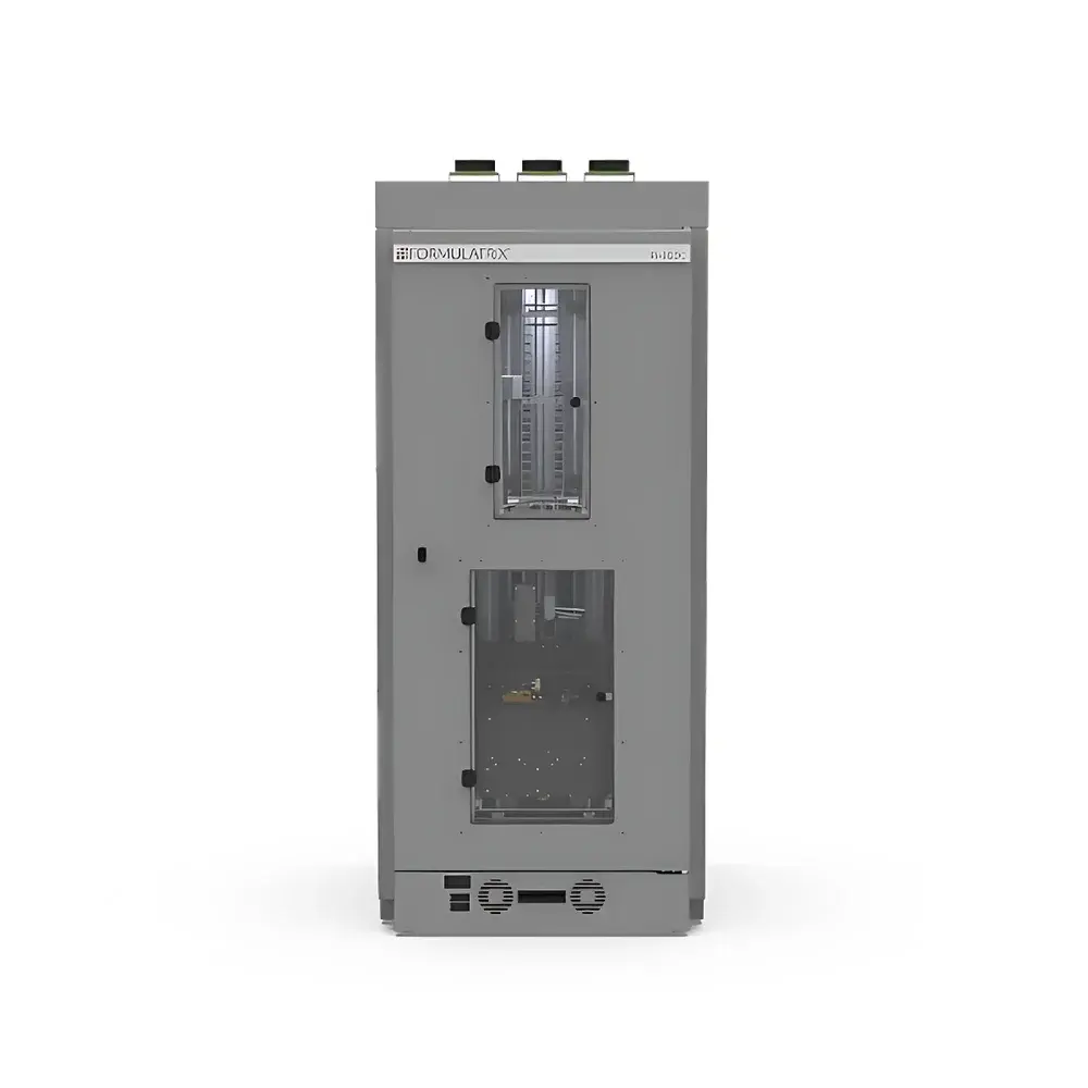
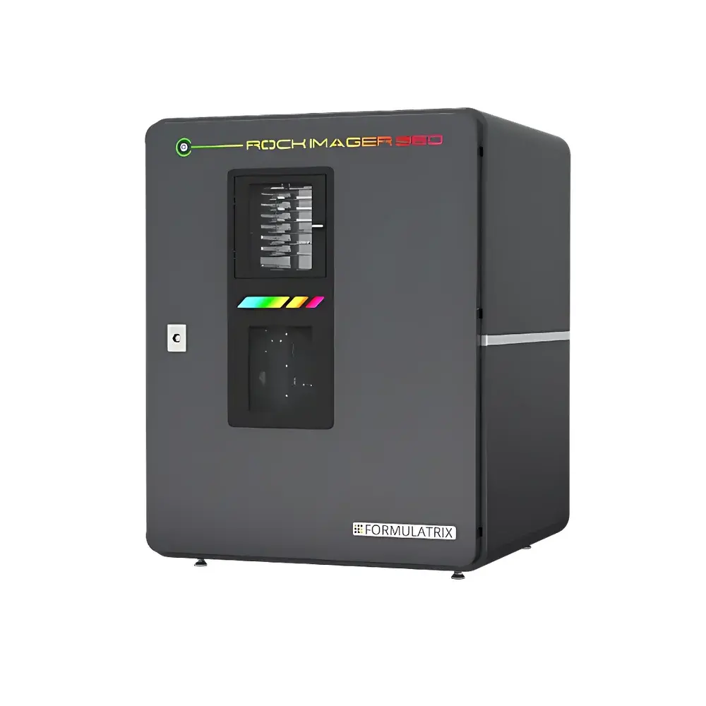
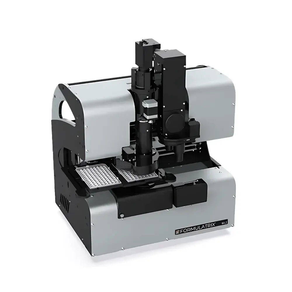
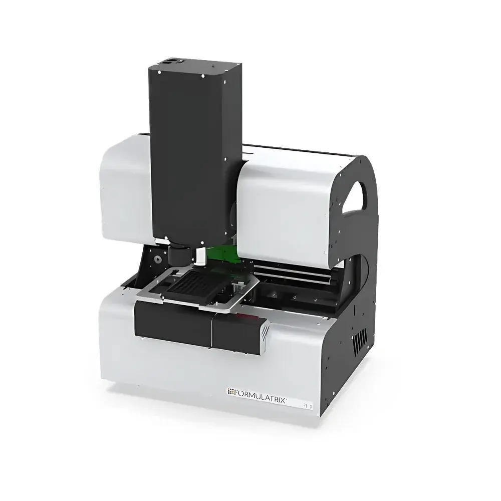
The Formulatrix Rock Imager RI360 and RI1000 are fully automated, high-precision protein crystal imaging systems engineered for structural biology laboratories engaged in high-throughput crystallization screening and optimization. Built on a robust robotic platform with integrated optical metrology, these instruments employ wide-field brightfield, polarized light, and UV fluorescence imaging modalities to capture morphologically and optically distinct features of protein crystals—particularly those embedded in challenging media such as lipidic cubic phase (LCP), microbatch, or vapor diffusion droplets. Unlike conventional static imagers, the Rock Imager implements real-time adaptive optics—including motorized focus stacking, dynamic exposure control, and intelligent droplet localization—to ensure reproducible, quantitative image acquisition across thousands of crystallization conditions without manual intervention. Its core architecture adheres to GLP-compliant design principles, supporting audit-trail-enabled operation under FDA 21 CFR Part 11–aligned software environments.
Key Features
- High-density plate handling: RI360 accommodates up to 364 SBS-standard microplates; RI1000 scales to 1,000 plates with programmable imaging schedules.
- Dual-modality optical path: Separately optimized visible-light and UV-fluorescence channels—UV channel features 280 nm LED excitation, UV-grade optics, and a 9.1 MP back-illuminated sCMOS sensor sensitive to tryptophan autofluorescence.
- Extended Focus Imaging (EFI): Combines high numerical aperture (NA) objectives with z-stack synthesis to maintain sub-micron resolution while extending effective depth of field—critical for imaging thick LCP slabs or multi-layered crystallization drops.
- Intelligent droplet localization: Proprietary computer vision algorithm detects and maps protein solution droplets—even within opaque or birefringent matrices—enabling ROI-based imaging without prior plate mapping.
- Precision thermal control: Peltier-based system maintains temperature between −5°C and +7°C relative to ambient, with stability ±0.5°C; RI1000 optional dual-compressor module extends range to 4–19°C with redundant cooling capacity.
- Automated optical calibration: Real-time adjustment of exposure time, polarization angle, iris aperture, and focus position per well—minimizing operator dependency and inter-plate variability.
Sample Compatibility & Compliance
The Rock Imager supports all major crystallization plate formats compliant with ANSI/SBS standards, including SBS 96-, 384-, and 1536-well plates, Linbro, Nextal, and custom LCP sandwich plates. Its non-contact imaging approach eliminates mechanical disturbance to fragile nucleation events. All hardware and firmware components meet IEC 61000-6-2/6-4 electromagnetic compatibility requirements. Data integrity is ensured via timestamped image metadata, hardware-logged environmental parameters (temperature, humidity, stage position), and optional 21 CFR Part 11–compliant electronic signatures when integrated with RockMaker or third-party LIMS platforms. The system is validated for use in ISO 17025-accredited labs and aligns with crystallization reporting guidelines from the Joint Center for Structural Genomics (JCSG) and the Structural Genomics Consortium (SGC).
Software & Data Management
Rock Imager operates natively via RockMaker—a Python-based application framework offering scriptable acquisition protocols, batch processing pipelines, and export to standardized formats (TIFF, HDF5, JSON). Image metadata conforms to the Crystallographic Information Framework (CIF)-compatible CrystalImageML schema. Integration with external platforms—including Mosflm, XDS, and Phenix—is supported through RESTful APIs and file-system watchers. Audit trails record every hardware command, parameter change, and user login event with SHA-256 hashing. Optional DICOM-SR export enables PACS integration for institutional biobanking workflows. All raw images retain full bit-depth (16-bit) and embedded calibration references for retrospective reprocessing.
Applications
- Primary crystallization screening across 96–1536-well plates with <3-minute acquisition per plate (visible light, fixed exposure).
- LCP-based membrane protein crystallization monitoring, including detection of sub-1 µm crystals obscured by lipid matrix using SONICC-ready parallel imaging modules.
- Quantitative FRAP analysis of fluorescently tagged proteins in crystallization drops to determine lateral diffusion coefficients and optimize lattice formation kinetics.
- Time-lapse morphological tracking of crystal growth, dissolution, or phase separation over days to weeks with zero operator presence.
- Validation of crystal identity against salt precipitates via birefringence contrast and SONICC second-harmonic generation signatures.
- Automated pre-screening for synchrotron beamline experiments—generating ranked lists of diffraction-quality candidates based on size, shape, and optical homogeneity metrics.
FAQ
What plate formats does the Rock Imager support?
SBS-compliant 96-, 384-, and 1536-well plates, Linbro, Nextal, and custom LCP sandwich plates—all recognized automatically via integrated barcode scanning.
Can the system distinguish protein crystals from salt crystals?
Yes—through combined analysis of birefringence, UV tryptophan fluorescence, and optional SONICC imaging, which detects non-centrosymmetric crystal lattices unique to proteins.
Is temperature control validated across the full operating range?
Yes—NIST-traceable PT100 sensors and independent thermographic verification confirm ±0.5°C accuracy from 4°C to 25°C.
How is data integrity maintained during long-term unattended operation?
Each image includes embedded EXIF metadata, SHA-256 checksums, and synchronized environmental logs; optional write-once archival storage prevents post-acquisition modification.
Does the system support remote monitoring and control?
Yes—via secure TLS-encrypted web interface with role-based access control, live camera preview, and queue status visualization from any modern browser.


