Grundium Ocus Digital Slide Scanner
| Brand | Grundium |
|---|---|
| Origin | Finland |
| Model | Ocus |
| Imaging Principle | High-Resolution Whole-Slide Scanning via Motorized Stage & Precision Optics |
| Objective Resolution | 0.48 µm/pixel (20× objective), 10 µm/pixel (1× overview lens) |
| Scan Area | User-Defined ROI (e.g., 15 mm × 15 mm in ~2 min) |
| Focus Mechanism | Fully Automated Z-Axis Autofocus + Software-Based Fine-Tuning & Manual Override |
| Stage | Motorized XY Stage Compatible with Standard 75 mm × 25 mm Microscope Slides |
| Onboard Compute | Integrated NVIDIA GPU-Accelerated Embedded PC |
| Storage | 500 GB Internal SSD, Expandable via USB 3.0 |
| Image Formats | TIFF, SVS, MRXS (Open Standard Compliant) |
| Connectivity | 1 GbE Ethernet, IEEE 802.11ac Wi-Fi, Offline Operation Supported |
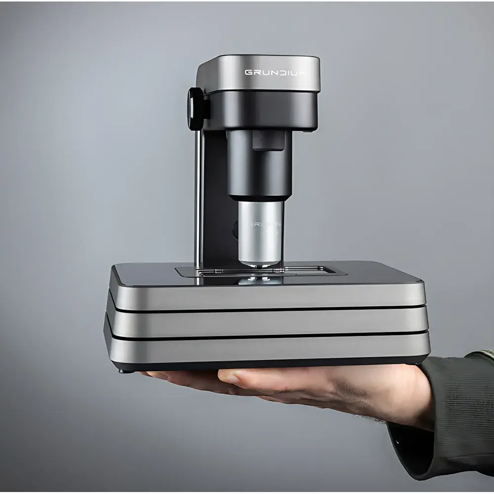
| Dimensions (W×D×H) | 180 × 180 × 190 mm |
| Weight | 3.5 kg |
| Illumination | Köhler-Optimized LED Light Engine |
| Sensor | 6 MP Monochrome/Color CMOS Sensor |
| UI Platform | Web-Based Interface (HTML5), Touchscreen-Compatible, Remote Access via Any Modern Browser |
| Compliance | Designed for GLP/GMP-Adjacent Workflows |
| Maintenance | Open-Chamber Design for Rapid Slide Loading and Optical Path Cleaning |
Overview
The Grundium Ocus Digital Slide Scanner is a compact, high-fidelity whole-slide imaging (WSI) system engineered for precision pathology digitization in resource-constrained or distributed laboratory environments. Unlike traditional benchtop scanners requiring dedicated server infrastructure, the Ocus integrates a GPU-accelerated embedded computer, Köhler-optimized LED illumination, and a calibrated motorized stage into a footprint smaller than an A4 sheet. It operates on a dual-optical-path architecture: a high-magnification 20× objective delivers 0.48 µm/pixel spatial resolution for diagnostic-grade cytological detail, while a 1× panoramic lens captures macroscopic context at 10 µm/pixel—enabling seamless navigation from tissue-level morphology to subcellular features. The system employs deterministic autofocus algorithms based on contrast gradient maximization across Z-stacks, ensuring reproducible focus acquisition even on uneven or thick-sectioned specimens. Its design prioritizes operational autonomy: all image acquisition, pyramid tiling, compression (lossless or JPEG2000), and metadata embedding occur onboard—eliminating dependency on external workstations or proprietary software licenses.
Key Features
- Self-contained imaging platform with integrated NVIDIA GPU and 500 GB SSD—no external PC required
- True whole-slide scanning with user-definable regions of interest (ROI), including rapid 15 mm × 15 mm scans completed in approximately 2 minutes
- Dual-resolution optical system: 20× objective (0.48 µm/pixel) for diagnostic analysis; 1× overview lens (10 µm/pixel) for orientation and triage
- Fully automated Z-axis autofocus with optional software-based fine-tuning or manual override during live imaging mode
- Web-native interface accessible via Chrome, Firefox, Safari, or Edge—supports touchscreens and remote access over LAN/WLAN without client installation
- Open hardware architecture: slide tray access, unobstructed optical path, and tool-free chamber cleaning facilitate routine maintenance and ISO 13485-aligned service protocols
- Compliance-ready output: generates multi-resolution SVS and MRXS files compatible with Qupath, QuPath, HALO, and commercial LIS/PACS systems supporting DICOM-SR extensions
Sample Compatibility & Compliance
The Ocus accepts standard 75 mm × 25 mm glass microscope slides with coverslip thicknesses ranging from 0.13–0.17 mm. Its mechanical stage accommodates both conventional H&E-stained sections and specialty preparations including IHC, FISH, and multiplex immunofluorescence—provided refractive index matching and mounting medium stability meet ISO 10993-5 cytotoxicity thresholds. While not FDA-cleared as a primary diagnostic device, the scanner’s output adheres to CAP checklist ANP.30710 (digital pathology validation) and supports laboratory-developed test (LDT) workflows under CLIA and ISO 15189 frameworks. Image metadata includes timestamp, objective ID, exposure parameters, and focus map coordinates—enabling audit trails required for GLP documentation and 21 CFR Part 11–aligned electronic records when deployed with validated identity management.
Software & Data Management
All image processing—including mosaic stitching, adaptive white balancing, color calibration against NIST-traceable standards, and pyramidal TIFF/SVS export—occurs locally within the embedded Linux environment. No cloud transmission is mandatory; data remains on-device unless explicitly exported via USB 3.0 or network share. The web-based viewer implements WSI-specific optimizations: progressive loading, region-of-interest prefetching, and GPU-accelerated rendering. Export formats comply with OpenSlide API specifications, ensuring interoperability with open-source analysis pipelines. For enterprise integration, RESTful APIs enable programmatic control of scan queues, metadata ingestion into LIMS, and automated QC flagging based on focus uniformity or signal-to-noise ratios.
Applications
- Remote frozen section consultation between satellite clinics and central pathology hubs
- Teaching archive creation with synchronized annotation layers for histology instruction
- Pre-analytical quality control in biobanking—rapid assessment of tissue integrity prior to molecular extraction
- Multi-center clinical trial slide digitization where data sovereignty and bandwidth limitations preclude centralized cloud upload
- Research-grade morphometric analysis using open-source tools such as Ilastik or CellProfiler, leveraging native TIFF exports with embedded scale bars
FAQ
Does the Ocus require a dedicated server or license subscription to operate?
No. All core functionality—including acquisition, processing, and viewing—is self-hosted on the device. No recurring software fees or cloud dependencies are involved.
Can the scanner be validated for use in a regulated diagnostic laboratory?
Yes. While the hardware itself is not IVD-certified, its deterministic scanning protocol, traceable calibration routines, and audit-log-capable firmware support IQ/OQ/PQ validation per ISO 13485 and CAP ANP.30710 guidelines.
What level of IT infrastructure is needed for network deployment?
Minimal. The device functions as a standalone HTTP server. Integration with existing Active Directory, LDAP, or PACS requires only standard network routing and firewall port configuration (TCP 80/443, 5000).
Is third-party image analysis supported?
Yes. Raw TIFF exports include EXIF-compliant spatial metadata. SVS and MRXS outputs are natively ingestible by QuPath, HALO, Visiopharm, and most FDA-cleared AI algorithms trained on TCGA or CAMELYON benchmarks.
How is long-term storage managed given the 500 GB internal drive?
Storage expansion is supported via USB 3.0 SSDs. Alternatively, scheduled rsync jobs or SMB/CIFS mounts can offload archives to NAS or object storage—without altering the scanner’s operational independence.
Related Products


