
Hiscore EOS Series High-Throughput Long-Term Live-Cell Real-Time Imaging and Analysis System
| Brand | Hiscore |
|---|---|
| Origin | Beijing, China |
| Manufacturer Type | Authorized Distributor |
| Region Classification | Domestic (China) |
| Model | EOS Series |
| Pricing | Available Upon Request |
Overview
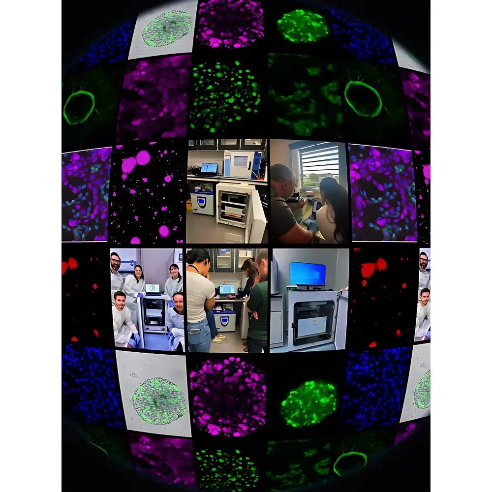
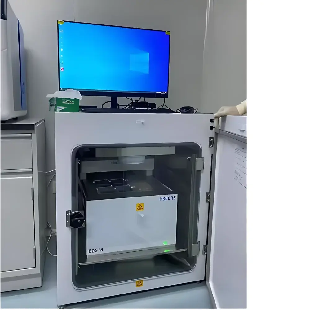
The Hiscore EOS Series is an engineered platform for high-fidelity, long-duration live-cell imaging and quantitative analysis under physiologically relevant conditions. Built upon a modular, incubator-integrated architecture, the system operates fully inside standard CO₂ incubators (37 °C, 5% CO₂, >95% humidity), eliminating thermal and environmental perturbations associated with sample retrieval. Unlike conventional inverted microscopes requiring external environmental chambers, the EOS embeds its optical train, motorized stage, and illumination subsystem directly within the incubator—enabling uninterrupted monitoring of cellular dynamics over periods exceeding 30 days. Its core imaging principle relies on widefield fluorescence and phase contrast microscopy, enhanced by precision Z-stack acquisition and AI-driven focus stabilization. Designed for reproducible longitudinal studies—including stem cell differentiation, organoid maturation, immunotherapy response tracking, and neurodevelopmental modeling—the EOS delivers spatially resolved, time-synchronized data streams compliant with GLP-aligned experimental workflows.
Key Features
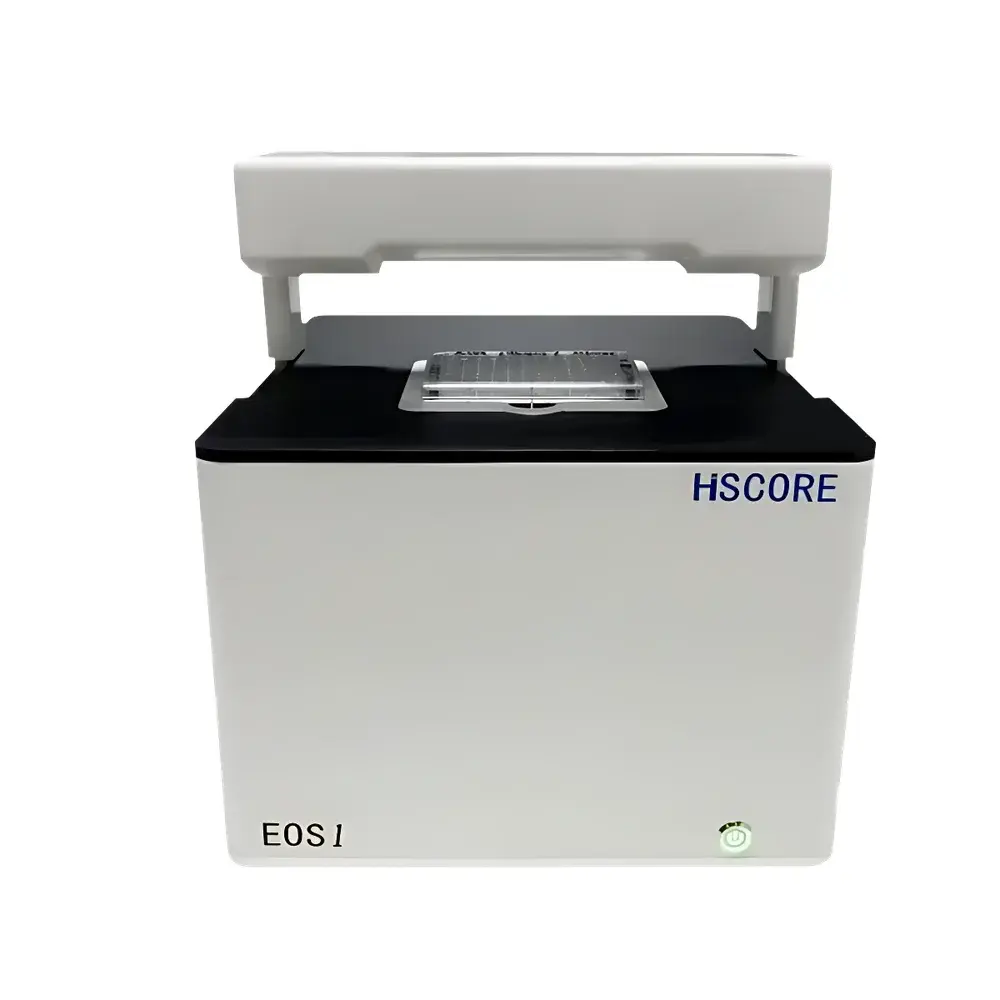
- Incubator-embedded design: Fully functional within standard cell culture incubators; no external temperature/humidity control required.
- Motorized 4-position objective turret: Supports simultaneous installation of Olympus-certified 4×, 10×, 20×, and 40× objectives; auto-switching enabled per protocol step.
- Multi-modal imaging: Brightfield, phase contrast, monochrome/multichannel fluorescence (405 nm, 488 nm, 561 nm, 640 nm, AO/PI), Z-stack, and whole-well imaging without stitching artifacts.
- AI-accelerated acquisition: Deep learning–based autofocus (DL-AF), exposure optimization, and real-time focus drift compensation ensure sub-micron stability across multi-day runs.
- Fixed-stage architecture: Sample plate remains stationary while optics move along X/Y/Z axes—minimizing mechanical disturbance to adherent or suspension cultures.
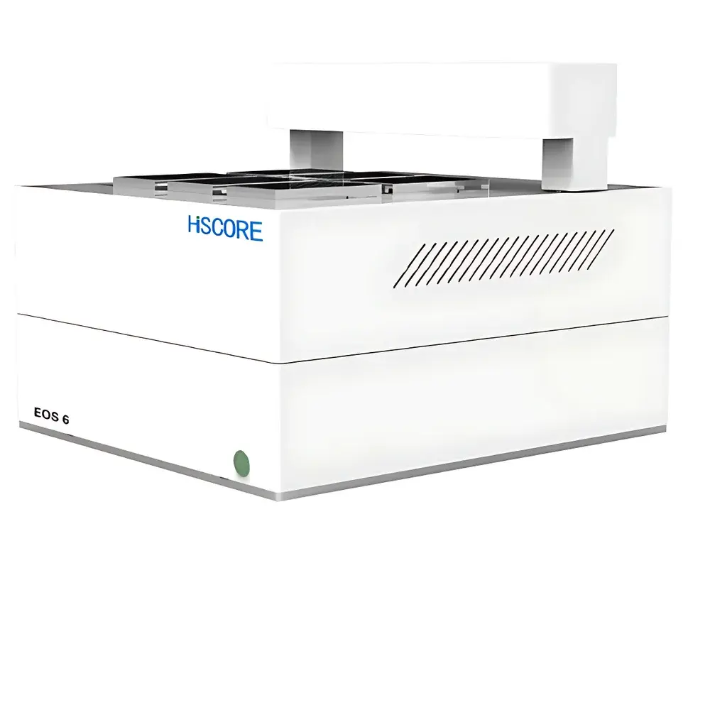
- High-throughput capacity: Up to six microplate positions (e.g., 6–384-well formats); independent channel selection, timing, and analysis per plate.
- Real-time output: 30 fps video recording (up to 180 s continuous), time-lapse image capture at user-defined intervals (seconds to hours), and single-frame acquisition.
Sample Compatibility & Compliance
The EOS accommodates both adherent and suspension cells—including primary isolates, iPSC-derived lines, spheroids, and organoids—without requiring surface coating or specialized substrates. It supports standard tissue-culture-treated plates, glass-bottom dishes, and hydrogel-embedded 3D matrices. All hardware components meet CE IVD and RoHS directives. Software complies with ALCOA+ data integrity principles and supports audit trail generation, electronic signatures, and role-based access control—facilitating alignment with FDA 21 CFR Part 11, ISO 13485, and GLP/GMP documentation requirements. Raw image metadata (timestamp, objective ID, exposure, z-position) is embedded in TIFF/OME-TIFF format per MIAME/MINSEQE standards.
Software & Data Management
The EOS Control Suite features a guided, icon-driven interface optimized for bench scientists—not software engineers. Integrated deep neural networks (U-Net variants trained on >10⁶ annotated cell images) perform pixel-level segmentation, lineage tracing, fluorescence intensity quantification, and morphometric classification. Analytical modules include confluence kinetics, proliferation/apoptosis profiling, phagocytosis scoring, cytotoxicity dose-response modeling (EC₅₀/IC₅₀ auto-calculation), neuronal arborization mapping, angiogenic sprouting quantification, and organoid growth trajectory analysis. Outputs include scatter plots, heatmaps, kinetic curves, and publication-ready TIFF/PDF reports. Data export supports HDF5, CSV, and JSON formats; API integration enables linkage with LIMS and ELN systems.
Applications
- Longitudinal phenotypic screening: Drug toxicity, compound efficacy, CRISPR knockout validation.
- 3D model dynamics: Organoid lumen formation, tumor spheroid invasion, vascular network remodeling.
- Immunology: T-cell–target cell interaction kinetics, macrophage phagocytic efficiency, NK-cell cytotoxicity assays.
- Neuroscience: Neurite outgrowth, synaptic puncta dynamics, calcium transient propagation in neuronal networks.
- Stem cell biology: Differentiation trajectory mapping, colony-forming unit (CFU) enumeration, pluripotency marker expression kinetics.
- Cell therapy QC: Viability, morphology, and functional consistency assessment pre- and post-cryopreservation.
FAQ
Can the EOS operate outside a CO₂ incubator?
No—the system is engineered exclusively for internal incubator deployment. Ambient operation compromises thermal/humidification stability and invalidates long-term viability claims.
Does the software support batch processing of historical datasets?
Yes. The analysis engine accepts imported TIFF stacks with metadata; reprocessing applies current AI models and calibration parameters retroactively.
Is Z-stack acquisition compatible with all fluorescence channels?
Yes. Multi-channel Z-stacks are acquired sequentially per plane with hardware-synchronized filter wheel and focus recalibration.
How is focus drift corrected during extended time-lapse sessions?
DL-AF performs continuous reference-layer correlation using low-magnification preview images, updating Z-position every 30–120 seconds based on real-time variance metrics.
What plate formats are supported for whole-well imaging?
6-, 12-, 24-, 48-, 96-, and 384-well plates; custom well geometry definitions can be imported via CSV coordinate files.


