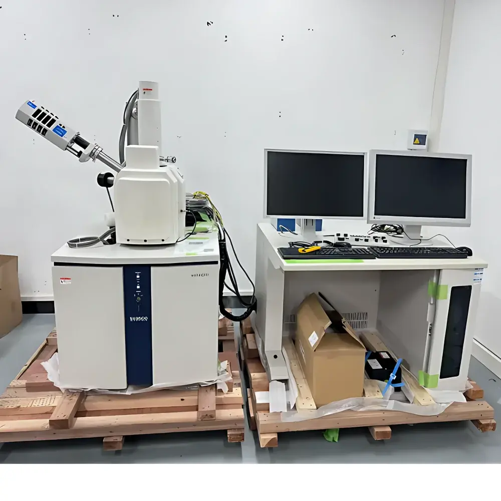
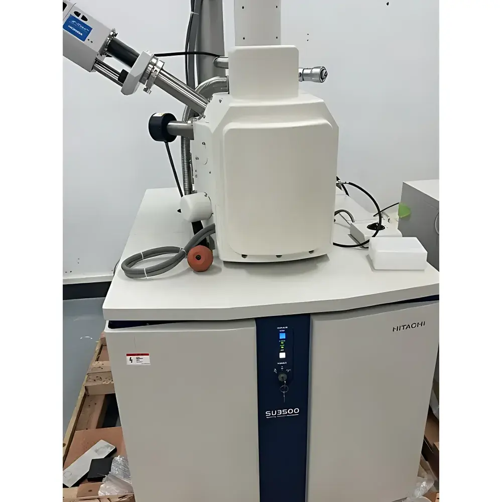
Hitachi SU-3500 Used Scanning Electron Microscope
| Brand | Hitachi High-Technologies |
|---|---|
| Origin | Japan |
| Manufacturer Type | Authorized Distributor |
| Category | Imported Instrument |
| Model | HITACHI SU-3500 |
| Price | USD 62,500 (FOB Yokohama) |
| Operational Age | 5 years |
| Secondary Electron Resolution | 3 nm @ 30 kV, 7.0 nm @ 3 kV |
| Backscattered Electron Resolution | 4.0 nm @ 30 kV, 10.0 nm @ 5 kV |
| Magnification Range | 5× to 300,000× (typical working range: 20×–50,000× for standard bulk samples) |
| Accelerating Voltage | 0.3–30 kV |
| Detector Configuration | In-lens SE detector, Everhart-Thornley SE detector, solid-state BSE detector |
Overview
The Hitachi SU-3500 is a field-emission-assisted, variable-pressure scanning electron microscope (SEM) engineered for high-resolution surface and cross-sectional imaging of non-magnetic solid materials. Designed with a thermionic tungsten filament electron source and optimized electromagnetic lens system, the SU-3500 delivers stable beam performance and reproducible imaging across a wide accelerating voltage range (0.3–30 kV). Its dual-mode detection architecture supports simultaneous acquisition of secondary electron (SE) and backscattered electron (BSE) signals—enabling topographic contrast, compositional contrast, and material phase differentiation without requiring sample coating in many cases. The instrument features a large chamber accommodating specimens up to Ø200 mm × 50 mm height, integrated stage tilt (±90°), and motorized X-Y-Z translation—making it suitable for routine QC inspection, failure analysis, and academic microstructural characterization.
Key Features
- High-stability thermionic electron source with extended filament lifetime and low beam drift
- Variable pressure mode (up to 133 Pa) for imaging of partially conductive or hydrated samples without full metal sputtering
- Dual-detector configuration: In-lens SE detector for ultra-high resolution at low kV; Everhart-Thornley SE detector for enhanced signal-to-noise ratio at higher magnifications; solid-state BSE detector for atomic number (Z)-contrast imaging
- Motorized 5-axis eucentric stage with ±90° tilt, enabling precise cross-sectioning alignment and angular view reconstruction
- Integrated digital image capture system with real-time frame averaging and dynamic focus correction
- Robust vacuum architecture featuring turbomolecular pump + rotary vane backing pump, achieving base pressure <5×10⁻³ Pa
Sample Compatibility & Compliance
The SU-3500 is validated for use with non-magnetic bulk solids—including metals (e.g., Al, Cu, Ti alloys), ceramics (Al₂O₃, SiC), geological specimens (rock thin sections, mineral grains), polymers, composites, fibers, and thin-film coatings. Samples must be electrically grounded or lightly carbon-coated for optimal charge dissipation. Magnetic specimens are excluded due to potential beam deflection and stage interference. The system complies with IEC 61000-6-3 (EMC emission standards) and meets mechanical safety requirements per ISO 13857. While not certified under FDA 21 CFR Part 11, its digital acquisition software supports audit-trail-enabled operation when configured with timestamped metadata logging—a prerequisite for GLP-compliant laboratories performing materials qualification per ASTM E1558 and ISO 16700.
Software & Data Management
Operation is managed via Hitachi’s proprietary SEM Control System (v4.2+), supporting intuitive point-and-click navigation, multi-region stitching, and automated focus/stigmation routines. Image data is saved in TIFF and BMP formats with embedded metadata (kV, WD, magnification, detector type, date/time stamp). Raw image files retain 16-bit grayscale depth for post-acquisition quantitative analysis using third-party tools (e.g., ImageJ, MountainsMap, or MATLAB-based particle analysis scripts). Export protocols support DICOM-compatible metadata tagging for integration into institutional LIMS environments. Remote diagnostics and firmware updates are available via secure SSH connection—subject to end-user network policy and Hitachi’s maintenance agreement terms.
Applications
- Microstructural analysis of sintered ceramics and refractory composites for porosity quantification and grain boundary delineation
- Failure analysis of fractured metallic components in aerospace and automotive QA/QC workflows
- Surface morphology assessment of catalyst supports (e.g., γ-Al₂O₃, zeolites) and nanocatalyst dispersion uniformity
- Cross-sectional imaging of multilayer thin-film stacks in photovoltaic R&D and semiconductor packaging evaluation
- Morphological characterization of electrospun polymer nanofibers and biodegradable scaffold matrices
- Mineralogical identification and textural mapping in geological core samples using BSE Z-contrast segmentation
FAQ
Is this unit refurbished or inspected prior to resale?
Yes—each SU-3500 undergoes full functional validation including vacuum integrity testing, electron optical alignment verification, detector sensitivity calibration, and stage repeatability assessment. A comprehensive inspection report and operational logbook are provided.
Does the system include EDX capability?
No—the listed configuration is SEM-only. An optional Bruker QUANTAX 200 or Thermo Scientific NSS energy-dispersive X-ray spectrometer may be integrated separately upon request, subject to chamber port availability and compatibility certification.
What service support is available post-purchase?
We provide 12 months of remote technical assistance and access to Hitachi-certified field engineers for on-site startup support. Extended warranty and preventive maintenance contracts are available directly through Hitachi High-Technologies’ global service network.
Can the instrument be shipped internationally with customs documentation?
Yes—full export compliance documentation (including ECCN classification, commercial invoice, packing list, and Certificate of Origin) is prepared in accordance with U.S. EAR and EU Dual-Use Regulation (EU No. 2021/821). Installation coordination includes pre-shipment electrical compatibility review (200–240 VAC, 50/60 Hz).
")