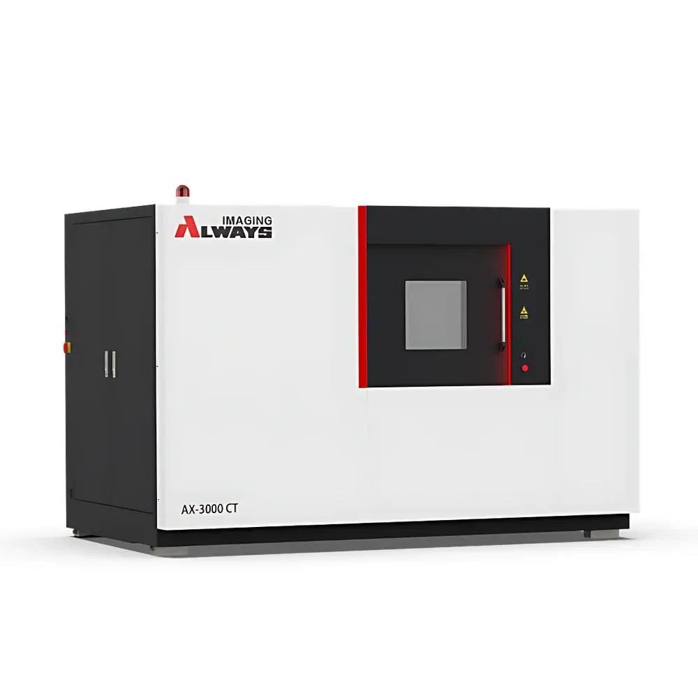
Always Imaging AX-3000CT Industrial Micro-CT System with Dual kV Options (225/240/300 kV) and Sub-2 µm Spatial Resolution
| Brand | Always Imaging |
|---|---|
| Origin | Shanghai, China |
| Manufacturer Type | Direct Manufacturer |
| Country of Origin | China |
| Model | AX-3000CT |
| Detector Type | Digital Flat-Panel Detector (139 µm pixel pitch, 3072 × 3072 pixels) |
| Scan Mode | Rotation-Only (RO) |
| Accuracy | Micron-Level |
| X-ray Energy | 225 kV / 240 kV / 300 kV |
| Penetration Capability | Sample-Dependent |
| Spatial Resolution | 1.5–2 µm (with optional 180 kV source down to 800 nm) / 3.5 µm |
| Density Resolution | <1% |
| Maximum Specimen Dimensions | Ø600 mm × H800 mm |
| Dimensional Measurement Accuracy | Sample-Dependent |
| Density Measurement Accuracy | Sample-Dependent |
| Max. Sample Weight | 25 kg / 50 kg / 100 kg |
| System Dimensions | 2950 × 1650 × 1950 mm |
| System Weight | 6000 kg |
| Radiation Shielding | Integrated Lead-Lined Enclosure (<1 µSv/h outside cabinet) |
| Cooling | Water-Cooled X-ray Source (500 W max power) |
| Focal Spot Size | 3–5 µm (standard), down to 800 nm (optional 180 kV reflective target) |
Overview
The Always Imaging AX-3000CT is a high-performance industrial micro-computed tomography (micro-CT) system engineered for non-destructive 3D internal and external metrology across diverse R&D and quality assurance environments. Operating on the principle of cone-beam X-ray computed tomography, the AX-3000CT acquires hundreds of projection images as the sample rotates 360° under a fixed microfocus X-ray source and digital flat-panel detector. Reconstruction algorithms (e.g., Feldkamp-Davis-Kress filtered backprojection) generate isotropic volumetric datasets with sub-micron spatial fidelity—enabling quantitative analysis of geometry, porosity, density distribution, material heterogeneity, and assembly integrity without physical sectioning. Its modular architecture supports dual-kV source configurations (225/240/300 kV), permitting optimized contrast-to-noise ratio for both low-Z polymers and high-Z metallic or geological specimens. The integrated lead-shielded enclosure complies with national radiation safety standards (e.g., GBZ 130-2020), ensuring operational safety in standard laboratory or production-floor settings.
Key Features
- Modular microfocus X-ray source platform: Selectable reflective or transmission targets; focal spot sizes from 3–5 µm (standard) to ≤800 nm (optional 180 kV configuration)
- Dual-voltage capability: Configurable at 225 kV, 240 kV, or 300 kV—enabling broad material penetration while preserving resolution for dense alloys, composites, and rock cores
- High-fidelity digital flat-panel detector: 3072 × 3072 active pixels, 139 µm pixel pitch, providing large field-of-view coverage (up to Ø600 mm × 800 mm height) without tiling artifacts
- Rotation-only (RO) scanning geometry: Minimizes mechanical complexity and vibration-induced blurring; ensures high reproducibility in repeated scans for longitudinal studies or statistical process control
- Integrated radiation shielding: Self-contained lead-lined cabinet meeting regulatory dose rate limits (<1 µSv/h at 5 cm from surface), eliminating need for external vault construction
- Scalable load capacity: Three mechanical stage options support samples up to 25 kg, 50 kg, or 100 kg—accommodating everything from MEMS devices to turbine blades and sedimentary core sections
- Water-cooled X-ray generator: Stable thermal management for continuous operation at up to 500 W output, critical for high-throughput inspection and time-resolved CT protocols
Sample Compatibility & Compliance
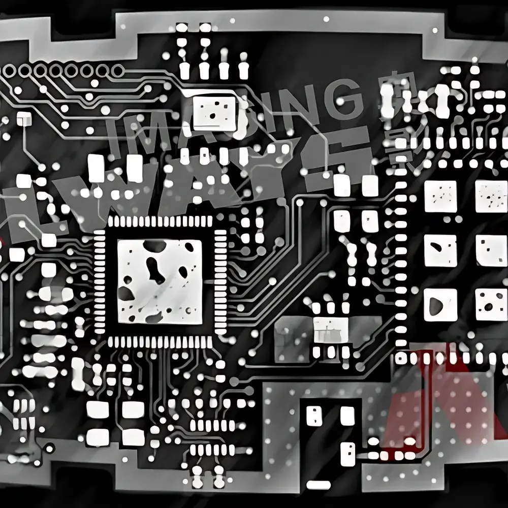
The AX-3000CT accommodates a wide spectrum of industrially and scientifically relevant specimens—including injection-molded polymer housings, die-cast aluminum components, PCB assemblies, semiconductor packages, fossilized biological structures, shale and sandstone core plugs, fiber-reinforced composites, battery electrode stacks, and hydrated soil aggregates. Its micron-level dimensional accuracy and <1% density resolution support compliance-driven workflows aligned with ASTM E1441 (Standard Guide for Computed Tomography), ISO 15732 (Non-destructive testing — Industrial computed tomography), and ASME BPVC Section V, Article 24. For regulated industries (aerospace, medical device manufacturing), raw projection data and reconstructed volumes retain full audit trails—compatible with GLP/GMP documentation frameworks and 21 CFR Part 11–compliant software extensions when deployed with validated acquisition modules.
Software & Data Management
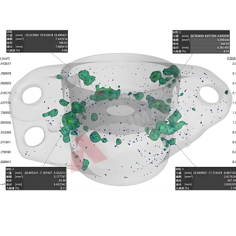
The system ships with proprietary reconstruction and analysis suite supporting GPU-accelerated FDK and iterative (SART, OS-SART) reconstruction, beam-hardening correction, ring artifact suppression, and phase-contrast enhancement where applicable. Volume rendering, multi-planar reformatting (MPR), and region-growing segmentation enable precise defect localization (e.g., voids, inclusions, delaminations). Metrology tools include GD&T evaluation (true position, cylindricity, profile), wall thickness mapping, and porosity quantification per ASTM E2957. All datasets are stored in DICOM 3.0 or NRRD format with embedded metadata (kV, µA, exposure time, rotation step, calibration coefficients). Export interfaces support Python API integration, MATLAB connectivity, and direct import into commercial CAE platforms (e.g., ANSYS, Thermo-Calc, Avizo).
Applications
- Aerospace & Defense: Wall thickness verification of turbine vanes; detection of casting porosity in Ti-6Al-4V structural components; analysis of thermal barrier coating integrity
- Electronics & Semiconductors: Void detection in solder joints; wire bond integrity assessment; package-level failure analysis (FA) of BGA and SiP assemblies
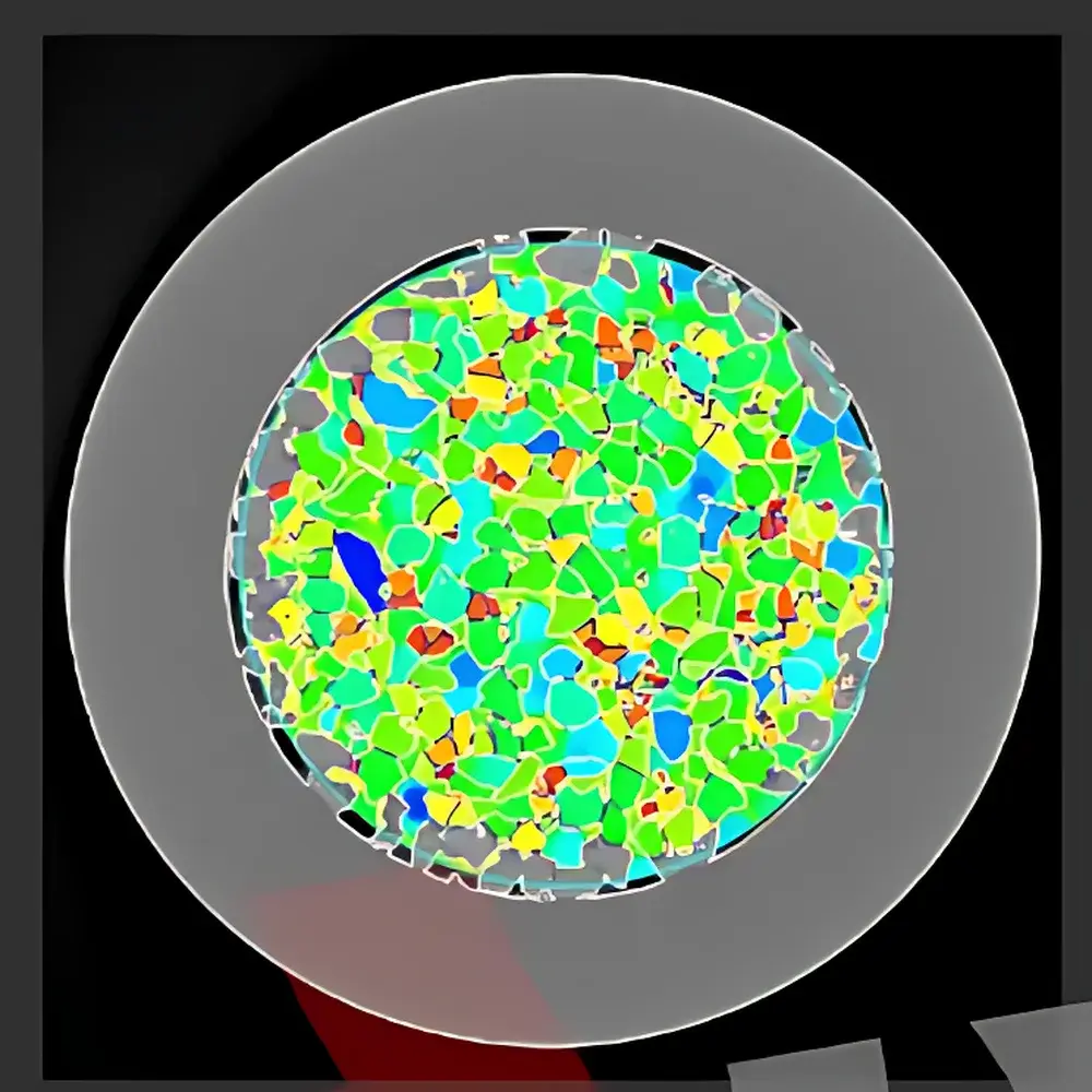
- Geosciences & Petroleum Engineering: Pore-network modeling of reservoir rocks; fluid saturation tracking in core flooding experiments; fracture aperture quantification
- Advanced Manufacturing: Validation of binder jetting and laser powder bed fusion AM parts; dimensional conformity checks against CAD models; residual stress-induced distortion mapping
- Life Sciences & Paleontology: Non-invasive morphometric analysis of fossilized vertebrate remains; trabecular bone architecture characterization; plant root-soil interface imaging
- New Energy: Electrode homogeneity evaluation in Li-ion cells; separator pore tortuosity measurement; degradation mechanism studies under cycling conditions
FAQ
What is the minimum achievable voxel size under optimal conditions?
With the optional 180 kV reflective target and high-magnification geometric setup, the AX-3000CT achieves effective isotropic voxel sizes down to 0.8 µm.
Can the system perform in-situ or time-lapse CT?
Yes—the RO scan mode, programmable stage control, and real-time projection streaming enable dynamic experiments such as thermal expansion monitoring, compression testing, and fluid infiltration studies.
Is the system compatible with third-party reconstruction software?
Raw projection data is exported in standard HDF5 or TIFF stack formats, fully interoperable with commercial (VGStudio MAX, Dragonfly) and open-source (Tomopy, ASTRA Toolbox) reconstruction pipelines.
Does the AX-3000CT support automated batch scanning?
The system includes scriptable acquisition protocols via Python SDK, enabling unattended multi-sample runs with pre-defined geometry, exposure, and reconstruction parameters.
How is radiation safety verified during commissioning?
A certified health physicist performs leakage and scatter surveys per IEC 61331-1 and local regulatory requirements; full test reports and dose mapping documentation are delivered with system handover.


