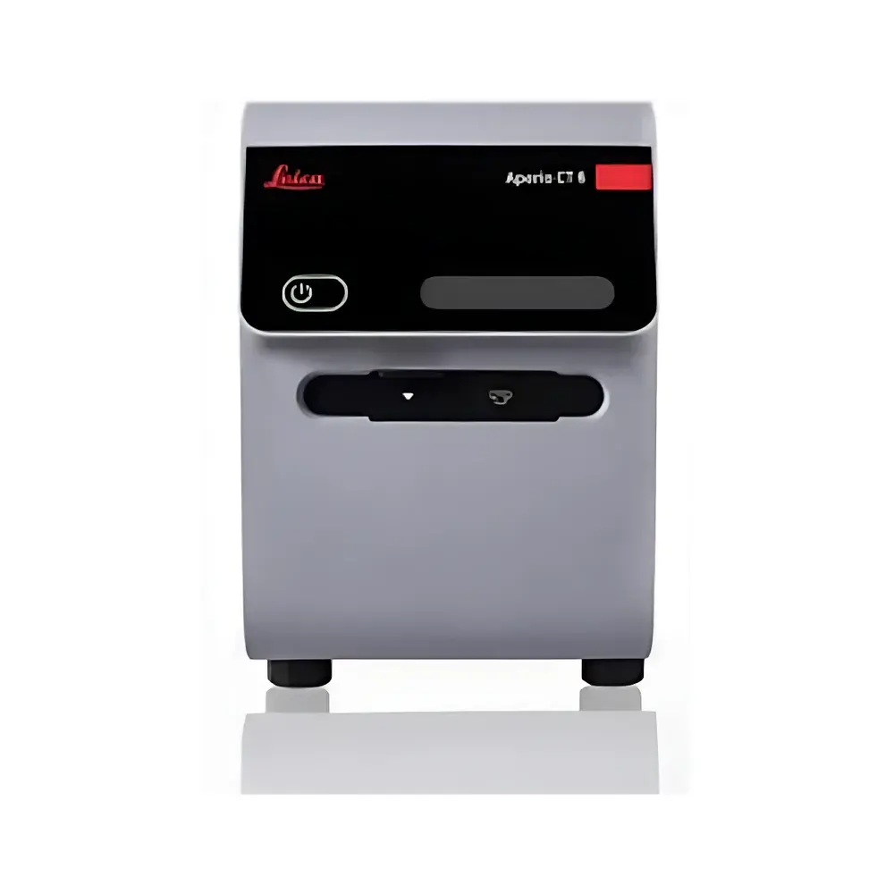
Leica Aperio CT6 Next-Generation Digital Pathology Desktop Scanner
| Brand | Leica |
|---|---|
| Origin | USA |
| Manufacturer Type | Authorized Distributor |
| Origin Category | Imported |
| Model | Aperio CT6 Next-Generation Digital Pathology Desktop Scanner |
| Pricing | Available Upon Request |
Overview
The Leica Aperio CT6 Next-Generation Digital Pathology Desktop Scanner is a high-performance, benchtop whole slide imaging (WSI) system engineered for precision, reproducibility, and seamless integration into modern pathology workflows. Built upon Leica Biosystems’ 150+ years of optical engineering excellence, the CT6 employs advanced line-scan technology—rather than conventional area-scan or tile-based acquisition—to deliver continuous, distortion-free image capture across glass slides. This optical architecture minimizes stitching artifacts, ensures pixel-perfect registration, and supports consistent focus depth control via dynamic Z-stack acquisition. Designed specifically for brightfield (BF) histopathology, the system captures high-resolution digital slides at up to 40× objective magnification (equivalent to 0.25 µm/pixel resolution), enabling diagnostic-grade review, quantitative image analysis, and long-term archival in compliance with CAP, CLIA, and ISO 15189 requirements.
Key Features
- True-color fidelity optics: Triple-prism RGB camera system with proprietary color calibration algorithms optimized for hematoxylin and eosin (H&E) staining—enhancing red-channel sensitivity to accurately distinguish erythrocytes, collagen, and nuclear chromatin contrast.
- Brightfield versatility: Native support for four standard brightfield stain types (H&E, IHC, Trichrome, PAS), with adaptive illumination profiling and stain-specific deconvolution modules for low-contrast or lightly stained sections.
- Flexible slide handling: Accommodates standard 1 × 3 inch (25 × 75 mm) microscope slides as well as extended formats up to 2 × 3 inches (50 × 75 mm); motorized stage enables rapid reconfiguration between routine and large-format scanning modes.
- Multi-layer focus fusion: Intelligent Z-stack acquisition with real-time focus mapping and algorithmic depth fusion—critical for uneven tissue sections, thick biopsies, or coverslip-variance scenarios—ensuring full-thickness morphological integrity in a single digital slide.
- Workflow adaptability: Dual-mode operation—fully automated batch scanning with barcode-driven slide identification, or manual single-slide acquisition with live focus preview and region-of-interest (ROI) selection—enabling both high-throughput labs and research-focused users to optimize throughput without compromising control.
Sample Compatibility & Compliance
The Aperio CT6 is validated for use with routinely processed formalin-fixed paraffin-embedded (FFPE) tissue sections, frozen sections, cytology preparations (e.g., Pap smears), and immunohistochemically stained specimens. It complies with key regulatory and quality frameworks including FDA-cleared software architecture (when deployed with Leica Biosystems’ certified Aperio eSlide Manager v13.x or later), HIPAA-compliant data handling protocols, and audit-trail functionality meeting 21 CFR Part 11 requirements for electronic records and signatures. All image metadata—including scanner model, objective lens ID, exposure parameters, color profile version, and user-defined annotations—is embedded in DICOM-SR and SVS file headers, ensuring traceability and interoperability with PACS, LIS, and AI-enabled analysis platforms.
Software & Data Management
The scanner operates natively with Leica Aperio ImageScope (viewer), Aperio eSlide Manager (enterprise-grade slide management), and Aperio Algorithms (quantitative pathology modules). All software components support DICOM WSI standards (Supplement 145), enabling vendor-neutral ingestion into hospital-wide imaging infrastructures. Integrated audit logging records every scan event—including operator ID, timestamp, slide barcode, acquisition settings, and post-processing actions—with immutable timestamps and role-based access controls. Data export options include SVS, TIFF, and DICOM WSI formats; encryption-at-rest and TLS 1.2+ transmission are enforced during network transfer to on-premise or cloud-hosted repositories.
Applications
- Clinical pathology: Primary diagnosis, second-opinion consultation, and telepathology services requiring CAP-accredited digital slide quality.
- Academic research: Longitudinal tissue studies, biomarker correlation, and training dataset generation for deep learning model development.
- Pharma & CROs: GLP-compliant digital archiving of toxicology and efficacy study slides, supporting FDA/EMA submission dossiers.
- Quality assurance: Routine QC of staining consistency, slide labeling accuracy, and scanner performance monitoring via built-in NIST-traceable reference slide calibration routines.
FAQ
Is the Aperio CT6 FDA-cleared for primary diagnostic use?
Yes—the system is FDA 510(k)-cleared (K221322) for primary diagnosis when used with Leica’s validated software stack and specified hardware configurations.
Does it support fluorescence or multiplex IHC scanning?
No—the CT6 is optimized exclusively for brightfield imaging; fluorescence and multispectral applications require the Leica Aperio GT 450 or Aperio AT2 platforms.
Can the CT6 integrate with existing LIS/PACS systems?
Yes—via DICOM WSI conformance and HL7 interface adapters, supporting bidirectional communication with major laboratory information systems and picture archiving systems.
What storage infrastructure is recommended for long-term slide archiving?
Leica recommends tiered storage solutions compliant with ISO/IEC 27001, including encrypted NAS with RAID 6 redundancy and automated migration to object storage (e.g., AWS S3 Glacier or Azure Archive Storage) for cold archive.
Is remote monitoring and maintenance supported?
Yes—Leica Remote Assist enables secure, consent-based technician access for firmware updates, calibration verification, and diagnostic log retrieval without local IT intervention.


