Leica Aperio GT450 Digital Pathology Scanner
| Brand | Leica |
|---|---|
| Origin | Germany |
| Model | Aperio GT450 |
| Scan Speed | 32 sec per 15 mm × 15 mm field at 40× magnification |
| Throughput | 81 slides/hour |
| Intended Use | For Research Use Only (RUO) |
| Optical System | Custom Leica 40× objective with real-time autofocus |
| Color Calibration | ICC-based medical-grade color correction |
| Image Quality Validation | Quadrant-based pathology review scoring (mean score > 3.75/4.0 across global pathology labs) |
Overview
The Leica Aperio GT450 Digital Pathology Scanner is a high-throughput, research-grade whole slide imaging (WSI) system engineered for reproducible, clinical-grade digital pathology workflows in academic, pharmaceutical, and translational research laboratories. Utilizing precision motorized stage control, synchronized LED illumination, and Leica’s proprietary optical architecture—including a custom-designed 40× apochromatic objective—the GT450 captures high-fidelity, focus-stable images across the entire tissue area via sequential tile acquisition and seamless stitching. Its core measurement principle relies on automated brightfield reflectance imaging with dynamic Z-stack optimization and real-time autofocus correction, ensuring consistent focal plane registration across heterogeneous tissue thicknesses and sectioning artifacts. Designed exclusively for research use (RUO), the system supports scalable deployment in GLP-aligned environments where traceability, repeatability, and image fidelity are foundational to downstream AI training, biomarker validation, and multicenter study harmonization.
Key Features
- High-speed scanning: Acquires a full 15 mm × 15 mm tissue region at 40× magnification in just 32 seconds—enabling rapid turnaround for large-volume slide cohorts.
- Continuous rack loading: Supports unattended operation with priority-based slide queuing and auto-feeding from standardized slide cassettes (up to 400 slides per batch).
- Real-time autofocus engine: Integrates Leica’s adaptive focus-tracking algorithm that dynamically adjusts Z-position during scanning—critical for maintaining optical clarity across variable tissue topography and coverslip thickness.
- Medical-grade color fidelity: Implements ICC v4-compliant color management with pre-calibrated display profiles, ensuring pixel-accurate chromatic reproduction aligned with conventional microscope viewing conditions (e.g., H&E, IHC, multiplex stains).
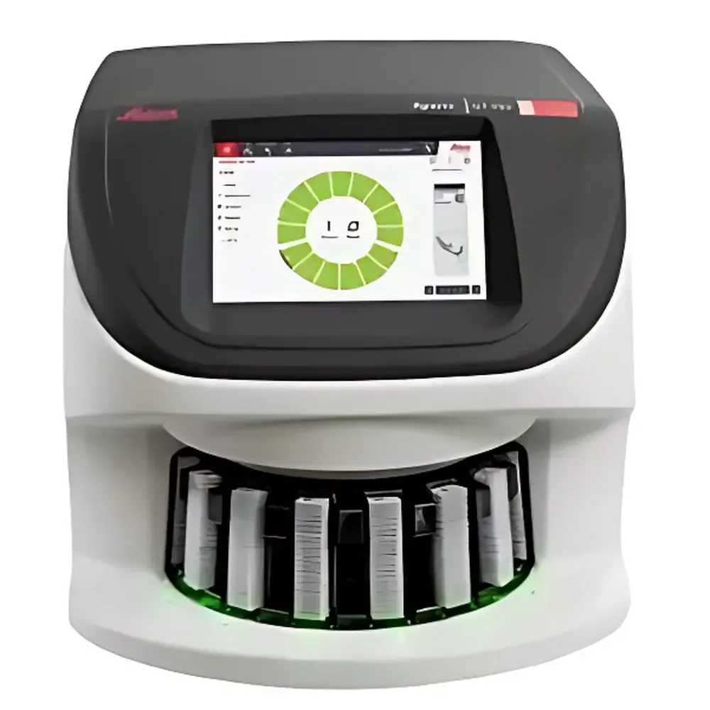
- Integrated touchscreen interface: Eliminates dependency on external PCs; all scan configuration, preview, and status monitoring performed directly on the device’s industrial-grade touch panel.
- Robust mechanical architecture: Built in Germany to ISO 9001-certified manufacturing standards, featuring vibration-damped optical bench, thermally stabilized LED illumination, and dust-resistant slide path design.
Sample Compatibility & Compliance
The Aperio GT450 accepts standard 1″ × 3″ glass microscope slides (75–110 µm thickness) with or without barcoded labels, compatible with routine formalin-fixed paraffin-embedded (FFPE) sections, frozen sections, cytology preparations, and tissue microarrays (TMAs). It supports common staining modalities including hematoxylin and eosin (H&E), immunohistochemistry (IHC), fluorescence in situ hybridization (FISH), and multiplex immunofluorescence (mIF) when used with appropriate filter sets (optional add-on). While designated RUO per regulatory classification, the system conforms to key international quality frameworks: its hardware design follows ISO 13485 principles for medical device-associated instrumentation, and software logs comply with ALCOA+ data integrity criteria. Audit trails, user authentication, and configurable retention policies support alignment with internal GLP protocols and FDA-aligned data governance practices for preclinical research.
Software & Data Management
The scanner operates natively with Aperio eSlide Manager™ v13.x or later—a DICOM-SUPP-compliant enterprise software platform supporting PACS integration, role-based access control, and encrypted DICOM WSI export (JPEG2000 or JPEG compression). All acquisitions generate pyramidal TIFF files compliant with OpenSlide and ASPIRE standards, enabling interoperability with third-party AI annotation tools (e.g., QuPath, HALO, Visiopharm). The embedded software includes built-in QC metrics—focus map heatmaps, stain uniformity histograms, and slide-level metadata tagging (stain type, date, operator ID)—all archived with SHA-256 checksums. Optional modules provide 21 CFR Part 11-ready electronic signatures, audit trail reporting, and automated backup to network-attached storage (NAS) or cloud object stores via S3-compatible APIs.
Applications
- Translational oncology research: Quantitative assessment of mitotic figures, nuclear morphology, and chromatin texture in breast cancer biopsies using AI-driven morphometric pipelines.
- Drug development histopathology: Centralized review of toxicologic endpoints across multisite preclinical studies, facilitating blinded peer consensus and inter-rater reliability analysis.
- Digital education & telepathology: Creation of annotated teaching libraries with embedded expert annotations, accessible via web-based viewers with pan/zoom/layer toggling.
- Biomarker discovery: High-resolution spatial profiling of protein co-expression patterns in tumor microenvironments using multiplex-stained serial sections.
- Quality assurance in core facilities: Standardized slide digitization protocols for reference slide archives, enabling longitudinal comparison and instrument performance trending over time.
FAQ
Is the Aperio GT450 cleared for clinical diagnostic use?
No—it is labeled For Research Use Only (RUO) and not intended for diagnostic procedures, patient management, or regulatory submission without additional validation per local jurisdiction.
Does the system support fluorescence scanning?
Fluorescence capability requires optional hardware modules (dual-band LED excitation, emission filter wheels, and sCMOS sensor upgrade); standard GT450 configuration is optimized for brightfield imaging only.
Can scan parameters be customized per slide batch?
Yes—users define per-batch protocols including magnification (20×, 40×), Z-stack depth, white balance presets, and region-of-interest (ROI) cropping masks via the touchscreen UI or eSlide Manager.
What file formats does it output?
Primary output is pyramidal TIFF (OpenSlide-compatible), with optional DICOM-SUPP export, JPEG2000 compression, and metadata-rich SVS header embedding.
How is image quality validated during installation?
Leica performs on-site IQ/OQ/PQ verification using NIST-traceable test slides and quadrant-based pathology review panels scored by certified pathologists against predefined morphological benchmarks.


