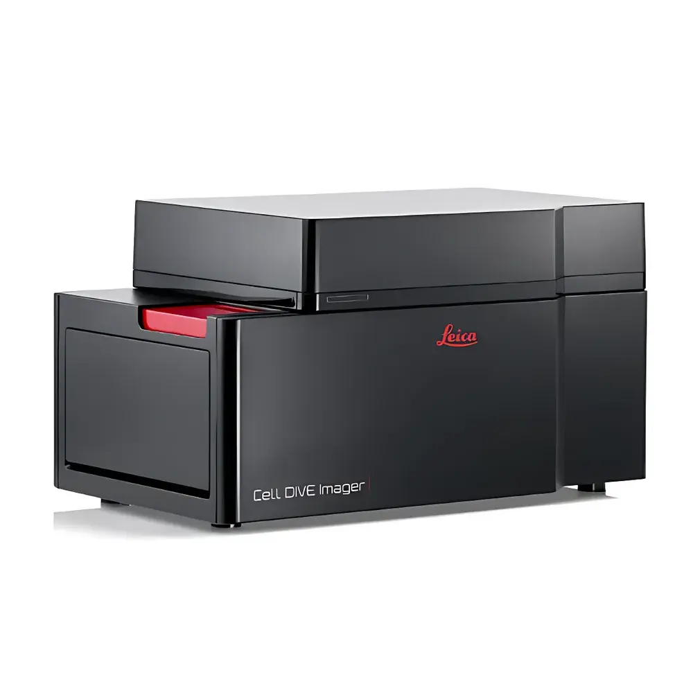
Leica CELL DIVE Ultra-Multiplex Tissue Imaging and Analysis System
| Brand | Leica |
|---|---|
| Origin | Germany |
| Model | CELL DIVE Solution |
| Category | Ultra-Multiplex Tissue Imaging & Spatial Biomarker Quantification System |
| Regulatory Status | CE-IVD compliant (for research use only in US) |
| Software Platform | CELL DIVE Analysis Suite v3.2+ |
| Imaging Modality | Cyclic Immunofluorescence (cycIF) with Automated Staining, Imaging, and Stripping |
| Max Biomarkers per Section | ≥60 |
| Spatial Resolution | Subcellular (≤300 nm lateral resolution with confocal mode) |
| Throughput | Up to 12 tissue sections per run |
| Data Output Format | OME-TIFF, JSON metadata, CSV cell-level feature tables |
| Compliance | ISO 13485 certified manufacturing, FDA 21 CFR Part 11–ready audit trail (software-enabled), GLP/GMP-compatible workflow logging |
Overview
The Leica CELL DIVE Ultra-Multiplex Tissue Imaging and Analysis System is a fully integrated, research-use-only platform engineered for spatially resolved, iterative immunofluorescence imaging of formalin-fixed paraffin-embedded (FFPE) and frozen tissue sections. It implements cyclic immunofluorescence (cycIF)—a rigorously validated methodology combining sequential antibody labeling, high-fidelity confocal or widefield imaging, and gentle, non-destructive fluorophore inactivation—to enable quantitative detection of ≥60 protein biomarkers on a single tissue section. Unlike conventional multiplex IHC (typically limited to 6–8 markers), CELL DIVE preserves tissue architecture and antigen integrity across repeated cycles, delivering single-cell–resolved spatial maps of cellular phenotypes, functional states, and intercellular neighborhood relationships. The system is purpose-built for translational oncology, immuno-oncology biomarker discovery, and spatial biology studies requiring statistically robust, reproducible quantification across large-cohort tissue microarrays (TMAs) or whole-section cohorts.
Key Features
- Automated cycIF Workflow: Integrated stainer-imager-stripper module enables unattended execution of up to 60+ antibody cycles with real-time focus maintenance, drift correction, and cycle-to-cycle registration accuracy ≤0.3 µm.
- Preserved Tissue Integrity: Proprietary mild stripping chemistry avoids epitope denaturation or section delamination—no antigen retrieval reapplication or physical section transfer required between cycles.
- Single-Cell Spatial Phenotyping: Combines subcellular-resolution imaging (300 nm lateral, 700 nm axial) with AI-assisted nuclear/cytoplasmic/membrane segmentation and morphology-aware cell typing.
- Validated Antibody Panel Ecosystem: Access to >250 rigorously pre-validated primary antibodies (human, mouse, rat), each tested for cross-reactivity, signal-to-noise ratio, and cycIF compatibility under standardized conditions.
- Hardware-Software Co-Optimization: Leica DMi8-based imaging engine with motorized stage, spectral unmixing-capable filter sets, and sCMOS camera (6.5 µm pixel size, 95% QE) synchronized to analysis software for traceable data provenance.
Sample Compatibility & Compliance
CELL DIVE supports standard FFPE sections (4–10 µm), OCT-embedded frozen sections, and cytospin preparations. All protocols adhere to ISO/IEC 17025–aligned validation frameworks. The system meets CE marking requirements under IVDR Annex II (Class B) for in vitro diagnostic research applications. For clinical trial support, workflows are compatible with CAP/CLIA laboratory documentation standards and support 21 CFR Part 11–compliant electronic signatures, audit trails, and user access controls when deployed with Leica’s validated IT infrastructure package. All reagents are supplied with CoA (Certificate of Analysis) and stability data per batch.
Software & Data Management
The CELL DIVE Analysis Suite (v3.2+) provides end-to-end computational processing: raw image alignment → spectral unmixing → cell segmentation (using U-Net trained on >10,000 annotated tissue regions) → marker intensity quantification → spatial statistics (e.g., nearest-neighbor distance, cellular co-localization index, neighborhood enrichment scoring). Data export conforms to OMERO and Bio-Formats standards (OME-TIFF), with metadata embedded per MIAME/MINSEQE guidelines. Batch processing pipelines are scriptable via Python API; all analysis parameters are version-controlled and reproducible. Audit logs record every user action, parameter change, and software update—fully traceable for GLP/GMP-aligned studies.
Applications
- Spatial characterization of tumor-immune interfaces in checkpoint inhibitor responders vs. non-responders
- Discovery of predictive cellular neighborhoods in early-stage NSCLC, melanoma, and CRC
- Longitudinal tracking of therapy-induced changes in stromal composition and immune cell infiltration
- Validation of digital pathology biomarkers against gold-standard manual scoring (inter-rater κ >0.85)
- Integration with bulk or single-cell RNA-seq datasets via spatial transcriptomics correlation modules
FAQ
How many cycles can be performed on a single tissue section without structural degradation?
Typical performance: ≥60 cycles with no measurable loss in section adhesion, nuclear morphology, or collagen autofluorescence baseline—validated across >1,200 independent FFPE samples.
Is antibody validation included with the system purchase?
Yes. Each instrument shipment includes access to the Leica Antibody Validation Portal, containing full validation reports (specificity, titration, cycIF performance) for all 250+ listed antibodies, plus protocol templates and troubleshooting guides.
Can CELL DIVE data be integrated into third-party analysis platforms like QuPath or HALO?
Yes. OME-TIFF exports are natively supported in QuPath v0.4+, HALO v3.4+, and Visium Spatial Gene Expression pipelines. Custom connectors for R/Bioconductor (spatialLIBD, Seurat v5 spatial modules) are available via GitHub repository.
What level of IT infrastructure is required for deployment?
Minimum: 64 GB RAM, dual Xeon Gold CPUs, NVIDIA A100 GPU (for accelerated segmentation), and 50 TB NAS storage with RAID 6 redundancy. Leica offers validated reference architectures and on-site IT readiness assessment.
Does the system support multiplexed DNA/RNA co-detection alongside protein markers?
Protein-centric design; however, optional integration with RNAscope HD duplex probes (ACD) is supported in hybrid workflows—requiring separate fixation optimization and validation per tissue type.
Related Products


