Leica DI C800 Digital Imaging Color Module
| Brand | Leica |
|---|---|
| Origin | Germany |
| Model | DI C800 |
| Component Type | Digital Imaging Module |
| Signal Input | XGA (1024 × 768) |
| Integration | Native Leica Microscope Interface |
| Application Domain | Surgical Microscopy, Ophthalmic Procedures |
Overview
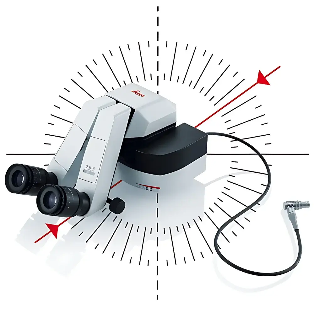
The Leica DI C800 Digital Imaging Color Module is a purpose-engineered optical imaging interface designed for seamless integration into Leica surgical microscopes—particularly those deployed in ophthalmic, neurosurgical, and microvascular applications. Operating on the principle of high-fidelity digital signal injection and real-time optical path overlay, the DI C800 accepts standard XGA-resolution (1024 × 768) video signals from external diagnostic or imaging sources—including fundus cameras, OCT systems, intraoperative ultrasound units, and digital slit lamps—and projects them directly into the surgeon’s binocular optical pathway. Unlike conventional external monitor-based visualization, this in-lens display architecture eliminates head movement away from the surgical field, preserving ergonomic posture, reducing visual accommodation latency, and maintaining continuous stereoscopic depth perception during critical interventions.
Key Features
- Native XGA (1024 × 768) digital signal compatibility with low-latency frame synchronization—optimized for real-time clinical decision-making.
- Optically aligned beam combiner module that integrates into Leica M841, M530, and FLX series microscope optical paths without compromising native magnification, resolution, or color fidelity.
- Zero-parallax overlay design ensures precise spatial registration between live surgical anatomy and superimposed diagnostic overlays (e.g., retinal layer segmentation maps or angiographic guidance markers).
- Compact, lightweight module (< 350 g) with passive thermal management—no active cooling fans or external power adapters required; powered via microscope’s internal bus interface.
- Adjustable brightness, contrast, and gamma correction accessible via dedicated rotary encoder—calibrated to meet ISO 13485-compliant ambient lighting conditions in Class I and II operating rooms.
- EMC-compliant construction per IEC 60601-1-2:2014, supporting safe co-location with electrosurgical units and MRI-adjacent environments.
Sample Compatibility & Compliance
The DI C800 is validated for use with Leica’s full line of modular surgical microscopes equipped with digital expansion ports (e.g., M841 Ergo, M530 OH6, FLX). It supports input from FDA-cleared and CE-marked diagnostic devices generating analog RGB or digital DVI-D (single-link) XGA output. The module itself carries CE marking under Directive 2017/745 (MDR), conforms to ISO 13485:2016 quality management requirements, and is classified as Class IIa medical device per Annex VIII of the MDR. Its optical performance meets EN ISO 10940:2020 standards for surgical microscope auxiliary displays, including luminance uniformity (>85%), chromaticity deviation (Δu’v’ < 0.005), and temporal response (< 25 ms rise/fall time).
Software & Data Management
The DI C800 operates as a hardware-level imaging conduit—not a standalone processing unit—and therefore requires no embedded firmware updates or proprietary software installation. All configuration parameters are stored in non-volatile memory within the microscope’s central control unit. When paired with Leica’s EnDoScan or ARTE software platforms, the module enables synchronized capture of dual-channel image streams: native ocular view + annotated diagnostic overlay. Audit trails—including timestamped activation logs, source device ID, and brightness settings—are recorded in accordance with ISO/TR 80001-2-2 for medical IT-network risk management and support GLP/GMP documentation workflows. No data storage occurs within the DI C800 itself; all images and metadata reside exclusively on the host microscope’s secure internal SSD or connected PACS infrastructure.
Applications
- Ophthalmic surgery: Real-time overlay of OCT B-scan cross-sections during vitrectomy or membrane peeling procedures.
- Neurosurgery: Registration of preoperative fMRI or DTI tractography data onto the operative field during tumor resection.
- Plastic and reconstructive surgery: Guidance via intraoperative fluorescence angiography (ICG) video feed projected directly into the eyepiece.
- ENT microsurgery: Synchronization of endoscopic sinus navigation data with microscopic anatomy during FESS procedures.
- Training and proctoring: Dual-stream recording capability supports objective assessment of resident hand–eye coordination and visual attention distribution.
FAQ
Does the DI C800 support HDMI or SDI inputs?
No—the module accepts only XGA-resolution analog RGB (with sync) or single-link DVI-D signals. HDMI and SDI require external format conversion compliant with IEC 62375-2:2017.
Can the DI C800 be retrofitted to older Leica microscope models?
Only models released after 2015 with the “Digital Expansion Port” (DEP) interface are compatible. Retrofitting to pre-2015 platforms (e.g., M320, M651) is not supported due to electrical and optical interface incompatibility.
Is calibration required before clinical use?
Yes—initial optical alignment and color balance calibration must be performed by an authorized Leica Field Service Engineer using Leica Calibration Suite v3.1 or later, per SOP-MICRO-DI-004.
Does the module introduce any measurable optical path length change?
No—the beam combiner introduces < 0.1 mm equivalent optical path difference, fully compensated within the microscope’s built-in tube lens system and verified during factory acceptance testing per ISO 10940 Annex D.
What is the expected service life under routine OR usage?
Rated for 30,000 hours of operational duty cycle at 23°C ambient; mean time between failures (MTBF) exceeds 120,000 hours per MIL-HDBK-217F predictions.


