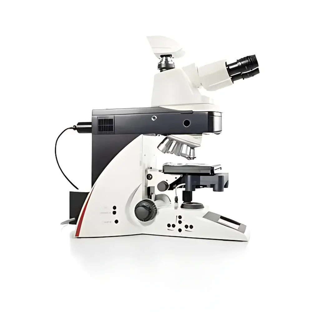
Leica DM4000 B LED Intelligent Biological Microscope with Integrated LED Illumination
| Brand | Leica |
|---|---|
| Origin | Germany |
| Model | Leica DM4000 B LED |
| Illumination Type | Integrated High-Stability White LED (50,000 h lifetime) |
| Illumination Modes | Brightfield, Darkfield, Phase Contrast, Polarization, Fluorescence |
| Objective Turret | Motorized 6-position or 7-position Smart Turret |
| Transmission Light Control | Fully Automatic Koehler Illumination with Objective Detection and Intensity Tracking |
| Fluorescence System | Motorized 5-position Filter Cube Selector, Motorized Field Diaphragm, Motorized Fluorescence Shutter, Zero-Drift Filter Positioning Technology |
| Fluorescence Intensity Management | Patented Fluorescence Intensity Management (FIM) with Channel-Specific Memory |
| User Interface | High-Resolution OLED Parameter Display |
| Ergonomic Controls | Six Programmable Thumb-Operated Function Keys on Focus Knob |
| Software Compatibility | Fully Compatible with Leica Application Suite (LAS X) and Third-Party CCD/CMOS Cameras (e.g., Leica DFC Series) |
Overview
The Leica DM4000 B LED is a high-performance, research-grade upright biological microscope engineered for precision life science applications—including cell biology, histopathology, developmental biology, and in vivo imaging workflows. Built upon Leica Microsystems’ decades of optical engineering excellence, the system employs advanced Köhler illumination principles for transmission light and optimized epifluorescence optics based on apochromatic correction. Its fully integrated, thermally stabilized white LED illumination source delivers consistent color temperature (≈5,700 K) across the full intensity range—eliminating spectral drift and enabling quantitative brightfield and fluorescence imaging without lamp warm-up or alignment. The microscope’s architecture supports multi-modal observation (brightfield, darkfield, phase contrast, polarization, and fluorescence) within a single platform, with automatic hardware reconfiguration triggered by user-selected imaging modes. Designed for reproducibility and compliance in regulated environments, the DM4000 B LED meets mechanical and optical stability requirements aligned with ISO 9001 manufacturing standards and supports audit-ready operation under GLP and GMP frameworks when paired with validated LAS X software.
Key Features
- Motorized 6- or 7-position smart objective turret with encoded position feedback for seamless mode switching and software synchronization.
- Fully automated transmission illumination system that detects objective magnification and NA in real time, dynamically adjusting LED intensity and aperture settings to maintain optimal Köhler alignment and signal-to-noise ratio.
- Patented Fluorescence Intensity Management (FIM): stores and recalls channel-specific excitation intensities, enabling rapid, repeatable, and phototoxicity-minimized multichannel acquisition.
- Zero-drift fluorescence filter cube positioning technology ensures sub-micron repeatability across sequential acquisitions—critical for time-lapse co-localization studies and quantitative ratiometric analysis.
- Integrated high-resolution OLED display showing real-time parameters: objective ID, magnification, condenser aperture, LED intensity (%), fluorescence filter position, shutter status, and focus position (when equipped with motorized Z-drive).
- Six programmable thumb-operated function keys mounted directly on the ergonomic focus knobs—enabling hands-on, eyes-on-sample operation without interface interruption.
- Modular design compliant with Leica’s HCX PL FLUOTAR and HCX PL APO objective series, supporting numerical apertures up to 1.40 and working distances optimized for live-cell imaging and thick-tissue sections.
Sample Compatibility & Compliance
The DM4000 B LED accommodates standard 26 mm and 30 mm coverslip thicknesses (0.13–0.17 mm), glass slides (1–1.2 mm), and specialized chambers including perfusion systems and stage-top incubators. It supports immersion media (oil, glycerol, water) via dedicated objective designs and maintains thermal stability during extended acquisition sessions. All optical components comply with ISO 8578 (microscope mechanical tube length) and DIN EN 61000-6-3 (EMC emissions). When operated with Leica LAS X software configured for audit trail logging and electronic signatures, the system satisfies FDA 21 CFR Part 11 requirements for data integrity in clinical diagnostics and pharmaceutical R&D. Routine calibration protocols align with ASTM E2877-22 (standard guide for microscope performance verification).
Software & Data Management
The microscope integrates natively with Leica Application Suite X (LAS X) v3.7+, offering full hardware control, multi-dimensional acquisition (Z-stacks, time-lapse, multi-channel tiling), and metadata embedding per frame (objective, illumination, exposure, gain, filter set). LAS X supports DICOM export, TIFF/OME-TIFF container formats, and direct linkage to laboratory information management systems (LIMS) via HL7 or RESTful APIs. Raw image data retains EXIF-compliant metadata, including illumination history and objective encoding—ensuring traceability for regulatory submissions. Optional LAS X Core modules enable automated cell counting, morphometric analysis, and fluorescence intensity quantification with built-in background subtraction and flat-field correction algorithms.
Applications
- Quantitative histopathology: standardized brightfield and IHC assessment using calibrated LED illumination and objective-matched contrast methods.
- Live-cell imaging: long-term observation with minimal phototoxicity via FIM-controlled LED excitation and environmental chamber compatibility.
- Fluorescence co-localization studies: zero-drift filter positioning enables pixel-accurate registration across multiple fluorophores over hours or days.
- Teaching and core facility deployment: intuitive one-button mode switching reduces training time while maintaining protocol fidelity across users.
- Regulated QC/QA labs: parameter logging, user authentication, and electronic record generation support ISO/IEC 17025 accreditation requirements.
FAQ
Does the DM4000 B LED support third-party cameras?
Yes—it features a standardized C-mount port and USB 3.0/USB-C interface for plug-and-play integration with scientific CMOS and CCD cameras from vendors including Hamamatsu, Andor, and Photometrics.
Is the LED illumination intensity adjustable in fine increments?
Yes—intensity is controllable in 0.1% steps across 0–100%, with hardware-level feedback ensuring linearity and stability independent of ambient temperature.
Can the system be validated for use in GxP environments?
When deployed with LAS X Pharma Edition and documented IQ/OQ protocols from Leica Service, the DM4000 B LED supports full GxP validation including calibration traceability to NIST standards.
What maintenance is required for the LED light source?
None—no alignment, no replacement, and no warm-up time. The LED module is rated for ≥50,000 operating hours at full output, with gradual lumen depreciation <3% per 10,000 hours.
How does the zero-drift filter technology improve multi-channel imaging?
It eliminates mechanical hysteresis and thermal drift in filter positioning, ensuring identical field-of-view registration across all channels—essential for accurate Pearson’s correlation coefficient calculation and spatial overlap analysis.


