Leica DMI3000 B Manual Inverted Microscope for Fundamental Life Science Research
| Brand | Leica |
|---|---|
| Origin | Germany |
| Model | DMI3000 B |
| Type | Manual Inverted Biological Microscope |
| Optical Configuration | Transmitted Light (Brightfield, Phase Contrast, DIC, Fluorescence) |
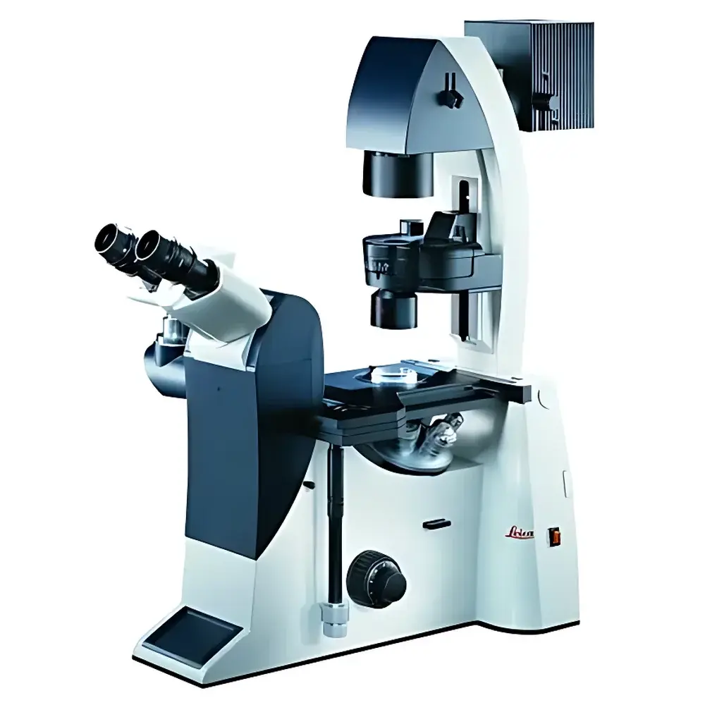
| Ergonomic Design | Adjustable Viewing Angle Binocular Tube with Central Viewing Port |
| Stability | High Thermal and Mechanical Stability |
| Intended Use | Live-cell Imaging, Time-lapse Microscopy, Micromanipulation, Routine Cell Culture Monitoring |
| Compliance | Designed to Support GLP/GMP-adjacent Lab Practices |
Overview
The Leica DMI3000 B is a manually operated inverted microscope engineered for reproducible, high-fidelity transmitted-light imaging in foundational life science laboratories. Its optical architecture is optimized for routine and advanced cell-based assays—including brightfield, phase contrast, differential interference contrast (DIC), and widefield fluorescence—without motorized automation or integrated digital capture. The system employs a fixed, high-stability mechanical platform with precision-ground optical pathways and thermally compensated components to minimize focus drift during extended time-lapse experiments. Unlike upright configurations, the inverted design positions the objective lenses beneath the specimen stage, enabling direct access to cultured cells in standard Petri dishes, flasks, and multi-well plates—critical for live-cell observation and micromanipulation workflows. The central viewing port between eyepieces ensures parfocal alignment across all magnifications and eliminates ocular positioning dependency, supporting consistent visual assessment regardless of operator height or seating posture.
Key Features
- Ergonomic binocular tube with continuously adjustable viewing angle (0°–35°), reducing cervical strain during prolonged use
- Central viewing port architecture: maintains optical path integrity independent of tube inclination
- Manual coarse/fine focusing mechanism with dual-speed coaxial knobs and 1 µm fine-focus graduation
- Stable inverted stand with reinforced cast-aluminum base and vibration-damped stage assembly
- Modular condenser system compatible with NA 0.3–0.9 dry condensers for phase contrast and DIC
- Dedicated fluorescence turret accommodating up to four filter cubes (e.g., DAPI/FITC/TRITC/Cy5), with manual cube selection
- Integrated halogen illumination (12 V/100 W) with field diaphragm, aperture diaphragm, and intensity control
- Stage with mechanical X-Y controls (76 × 52 mm travel), calibrated scale, and optional specimen holder adapters
Sample Compatibility & Compliance
The DMI3000 B accommodates standard tissue culture vessels including 35 mm–150 mm Petri dishes, T-25 to T-225 flasks, 6–96-well plates, and chambered coverglasses—enabling direct observation of adherent and suspension cultures without transfer. Its open-stage design permits integration with external micromanipulators, patch-clamp rigs, or environmental chambers (CO₂, temperature, humidity control). While the instrument itself does not carry FDA 510(k) or CE-IVD certification as a diagnostic device, its optical performance and mechanical consistency align with method validation requirements under ISO 13485-supported quality systems. Laboratories performing GLP-compliant cell line characterization or GMP-adjacent bioprocess monitoring routinely deploy the DMI3000 B as a reference imaging tool, supported by documented calibration procedures for focus repeatability and illumination uniformity.
Software & Data Management
As a manual platform, the DMI3000 B does not include embedded imaging software or onboard storage. However, it is fully compatible with third-party digital camera systems (e.g., Leica DFC series, Hamamatsu ORCA, or Point Grey/FLIR models) via C-mount or proprietary adapter interfaces. When coupled with acquisition software such as Leica Application Suite (LAS X) Lite, NIS-Elements, or open-source platforms like Micro-Manager, users can perform time-lapse capture, Z-stack acquisition, and basic fluorescence intensity quantification. All image metadata—including objective magnification, filter set, exposure time, and stage coordinates—is preserved in TIFF or OME-TIFF formats. Audit trail functionality depends on the selected host software; LAS X supports user-defined annotation fields and exportable acquisition logs compliant with 21 CFR Part 11 when deployed on validated Windows workstations.
Applications
- Routine morphology assessment of mammalian, insect, and stem cell lines during passaging and confluency monitoring
- Phase contrast-based viability estimation without staining (e.g., trypan blue exclusion correlation)
- Time-lapse documentation of mitosis, migration, wound healing, and neurite outgrowth over hours to days
- Fluorescent protein expression tracking (e.g., GFP-tagged cytoskeletal elements) using narrow-band excitation
- Manual microinjection, cell sorting, and patch-clamp electrode positioning under high-magnification DIC
- Quality control of primary cell isolates and organoid formation in developmental biology studies
FAQ
Is the DMI3000 B suitable for quantitative fluorescence measurements?
It supports semi-quantitative widefield fluorescence but lacks built-in intensity calibration or spectral deconvolution. For rigorous quantification, users must implement external reference standards, flat-field correction, and controlled exposure normalization.
Can this microscope be upgraded to motorized focusing or automated stage control?
No—the DMI3000 B is a fixed-function manual platform. Leica’s DMI6000/DMI8 series offer modular motorization, but retrofitting is not supported.
What maintenance intervals are recommended for long-term optical stability?
Annual verification of Köhler illumination alignment, condenser centering, and focus mechanism backlash is advised. Objective cleaning should follow ISO 10110-7 protocols using lens-grade solvents and lint-free wipes.
Does the system comply with ISO/IEC 17025 for testing laboratory accreditation?
While not certified per se, its mechanical and optical specifications meet traceable metrology benchmarks required for method validation within accredited labs—provided operational procedures and calibration records are maintained per ISO/IEC 17025 Clause 6.4.
Related Products


