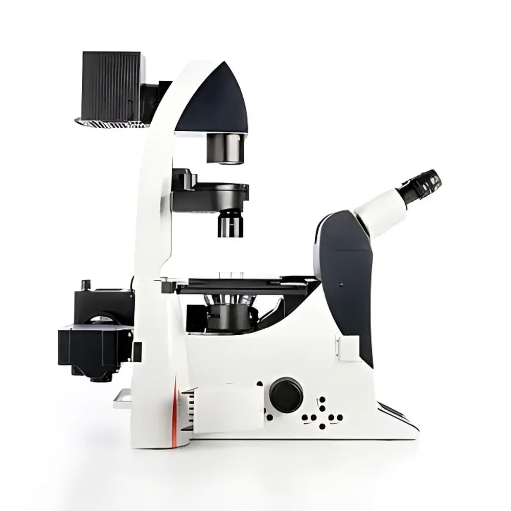
Leica DMI6000 B Automated Inverted Microscope for Biomedical Research
| Brand | Leica |
|---|---|
| Origin | Germany |
| Model | DMI6000 B |
| Type | Motorized Inverted Research Microscope |
| Optical Configuration | Dual-Path DIC/Fluorescence Compatible |
| Focus Drive | Motorized Z-axis with Piezo-assisted Fine Focus |
| Tube Options | Ergonomic Tilting Trinocular Tube (100% / 50% Beam Split) |
| DIC Components | Interchangeable Wollaston Prisms for Thin/Thick Specimens |
| Illumination Control | Automated Light Intensity, Field Diaphragm & Aperture Diaphragm Management |
| Compliance | Designed for GLP/GMP-aligned workflows |
Overview
The Leica DMI6000 B is a fully motorized inverted research microscope engineered for high-precision, reproducible imaging in demanding biomedical applications—including live-cell time-lapse microscopy, multi-channel fluorescence tomography, micromanipulation, and quantitative phase-contrast analysis. Its optical architecture integrates advanced Köhler illumination, high-numerical-aperture objective turrets, and dual-path beam-splitting capability to support simultaneous or sequential DIC (Differential Interference Contrast) and fluorescence modalities without mechanical realignment. The system operates on the principle of shear-based optical path difference detection in DIC, where minute refractive index gradients across biological specimens are translated into high-contrast, pseudo-3D relief images—enabling visualization of unstained, living cells with sub-micron structural fidelity. As an inverted platform, the DMI6000 B places the objective beneath the sample stage, optimizing accessibility for culture vessels, microfluidic chips, and electrophysiology rigs while maintaining thermal and mechanical stability during long-duration acquisitions.
Key Features
- Fully motorized optical train with programmable Z-axis drive (step resolution ≤ 10 nm) and piezo-enhanced fine focus for sub-nanometer axial positioning repeatability
- Dual-path DIC/fluorescence management system allowing independent control of both optical paths—enabling seamless switching between DIC contrast enhancement and multi-spectral fluorescence detection
- Ergonomic tilting trinocular tube (adjustable 0–35°) with configurable beam splitting (100% camera / 50:50 camera:eyepiece) and optional Bertrand lens integration for precise Köhler alignment and phase ring centering
- Modular Wollaston prism turret supporting application-specific shear optimization: low-shear prisms for thick tissue sections (>50 µm), high-shear variants for subcellular membrane dynamics in monolayer cultures
- Automated light management suite including intensity-regulated LED or metal-halide illumination, motorized field diaphragm, and aperture diaphragm—calibrated via hardware-level feedback loops to maintain constant photon flux across magnifications and objectives
- Integrated autofocus system with contrast-based and reflection-based algorithms, validated for glass-bottom dishes, polymer substrates, and hydrogel-embedded samples
Sample Compatibility & Compliance
The DMI6000 B accommodates standard and custom specimen formats: 35 mm–150 mm Petri dishes, multi-well plates (6–384-well), chambered coverglasses, microfluidic PDMS devices, and slice culture inserts. Its stage design supports temperature-controlled environmental chambers (20–40 °C), CO2 regulation modules (5% ± 0.2%), and humidity stabilization units—ensuring physiological relevance during extended live imaging sessions. From a regulatory perspective, the platform’s deterministic hardware control logic, audit-trail-capable firmware, and compatibility with Leica LAS X software (validated for 21 CFR Part 11 compliance) make it suitable for preclinical assay development under GLP conditions. It meets ISO 9001 manufacturing standards and conforms to IEC 61000-6-3 (EMC) and IEC 61010-1 (safety) requirements for laboratory instrumentation.
Software & Data Management
Control and acquisition are managed through Leica LAS X software—a modular, scriptable platform supporting automated multi-dimensional acquisition (XYZTλ), spectral unmixing, deconvolution, and quantitative intensity profiling. LAS X includes built-in tools for DIC vector calibration, fluorescence bleed-through correction, and Z-stack reconstruction with sub-pixel registration. Raw image data is stored in TIFF or Leica’s proprietary LIF format, preserving metadata such as objective ID, exposure parameters, DIC prism position, and stage coordinates. Export pipelines support standardized formats (OME-TIFF, ND2, CZI) for interoperability with ImageJ/Fiji, Imaris, and commercial AI-based segmentation suites. Audit trails log all user actions, parameter changes, and instrument state transitions—enabling full traceability required in regulated environments.
Applications
- Long-term live-cell tracking of mitotic progression, organelle motility, and cytoskeletal remodeling under physiological conditions
- Multi-parameter phenotypic screening in drug discovery workflows—combining DIC morphology assessment with calcium, pH, or ROS fluorescent reporters
- High-content analysis of stem cell differentiation kinetics using label-free DIC texture metrics alongside nuclear translocation markers
- Microinjection and patch-clamp experiments requiring simultaneous visual guidance (DIC) and fluorescence monitoring of ion-sensitive dyes
- 3D reconstruction of organoid architecture via automated Z-series acquisition with adaptive focus correction at each plane
FAQ
Is the DMI6000 B compatible with third-party micromanipulators and electrophysiology rigs?
Yes—the microscope features standardized mounting interfaces (M6/M4 threads) and TTL/RS-232 connectivity for synchronized triggering with external hardware including Eppendorf TransferMan, Sutter MP-285, and Axon Instruments systems.
Can DIC and fluorescence channels be acquired simultaneously without optical crosstalk?
The dual-path optical design enables true parallel acquisition; however, optimal separation requires appropriate dichroic mirrors and emission filters matched to the selected fluorophores and Wollaston prism orientation.
Does the system support automated Z-stack acquisition with drift correction?
Yes—LAS X implements both hardware-based (motorized stage + piezo Z-drive) and software-based (cross-correlation registration) drift compensation during time-lapse Z-series collection.
What level of validation documentation is available for regulated laboratories?
Leica provides IQ/OQ protocols, installation checklists, and firmware version traceability reports; full 21 CFR Part 11 validation packages are available upon request through certified service partners.
Are replacement Wollaston prisms calibrated per objective magnification?
Each prism is pre-aligned for a specific objective range (e.g., 10×–40× or 63×–100×); recalibration is performed automatically via the DIC manager when changing objectives or prisms.
Related Products


