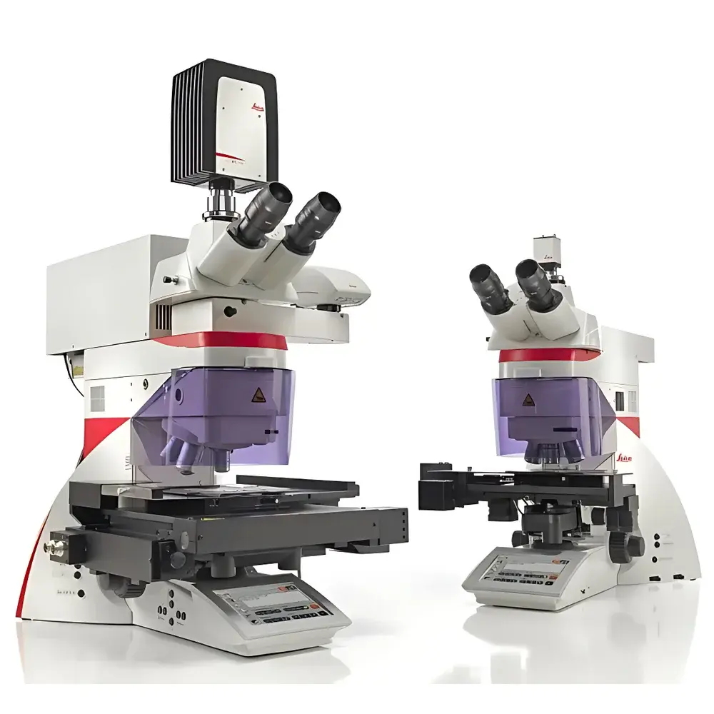
Leica LMD6 Laser Microdissection System
| Brand | Leica |
|---|---|
| Origin | Germany |
| Model | LMD6 |
| Cutting Principle | Gravity-assisted, beam-scanning laser microdissection |
| Optical Architecture | Upright microscope platform with galvanometric mirror-based laser beam steering |
| Sample Collection Method | Gravity-driven direct drop into collection vessels (e.g., PCR caps, 8-strip tubes, ibidi µ-slides, PEN-membrane slides) |
| Software Platform | Leica Application Suite (LAS) X v7.6 with AVC (Automatic Cell Recognition), time-lapse imaging, and workflow-driven ROI selection |
| Compliance | Designed for GLP/GMP-aligned workflows |
| Slide Compatibility | PEN membrane slides, PET slides (low-additive), DIRECTOR slides (film-free), standard glass slides |
Overview
The Leica LMD6 Laser Microdissection System is a high-precision, upright-platform optical instrument engineered for contact-free, contamination-free isolation of morphologically defined regions from heterogeneous tissue sections or cultured cell monolayers. Unlike conventional stage-scanning microdissection platforms, the LMD6 employs a fixed-sample, dynamic-beam architecture: a UV laser (355 nm) is precisely steered across the specimen plane using high-stability galvanometric mirrors and optimized achromatic optics—enabling true vertical-incidence cutting with minimal thermal spread and zero mechanical stress. This beam-scanning principle eliminates stage vibration, positional drift, and sample deformation, ensuring micron-level spatial fidelity and reproducible cut geometry across serial sections. The system operates exclusively under gravity-based collection: dissected material detaches cleanly and drops directly into user-selected receptacles—including PCR tube caps, 8-well strip tubes, ibidi µ-Slides, or PEN/PET-coated slides—without fluid transfer, enzymatic treatment, or adhesive contact. This design satisfies stringent preanalytical requirements for downstream omics applications where molecular integrity must be preserved.
Key Features
- Fixed-sample, scanning-beam architecture with UV laser (355 nm) and diffraction-limited spot size for clean ablation and minimal collateral damage
- Integrated upright microscope platform compatible with standard histological slides, live-cell culture dishes, and climate-controlled chambers (optional)
- Real-time “Move-Cut” mode enabling direct, operator-guided laser path definition via mouse or multi-touch interface
- Gravity-driven collection ensures 100% contactless transfer—no tape, no capillary action, no solvent exposure
- Leica Application Suite (LAS) X v7.6 software with modular add-ons: Automatic Cell Recognition (AVC), time-lapse acquisition, ROI database management, and customizable annotation layers
- Comprehensive slide compatibility: PEN membrane slides (genomics/transcriptomics), low-additive PET slides (proteomics/metabolomics), and film-free DIRECTOR slides for additive-sensitive workflows
Sample Compatibility & Compliance
The LMD6 accommodates both fixed and live specimens without hardware modification. Frozen or FFPE tissue sections (4–20 µm), cytospins, blood smears, and adherent cell cultures—including primary neurons, stem cell colonies, and organoid monolayers—are routinely processed. For live-cell applications, optional environmental control modules maintain CO₂, humidity, and temperature during dissection. All collection workflows comply with ISO 15189 preanalytical guidelines and support alignment with CLIA, CAP, and GCP frameworks. When configured with secure user authentication, electronic signatures, and enabled audit trail logging, the LAS X software meets documentation requirements for FDA 21 CFR Part 11 compliance in regulated environments. No consumables requiring lot traceability (e.g., tapes or adhesives) are used—reducing variability in nucleic acid yield and protein recovery.
Software & Data Management
LAS X v7.6 provides a unified, workflow-oriented interface built on a modular architecture. Users define ROIs via freehand drawing, polygonal selection, or intensity-threshold segmentation. AVC module enables unsupervised identification of morphologically distinct cell populations based on texture, size, and nuclear/cytoplasmic contrast—exportable as binary masks for batch processing. Time-lapse imaging captures dynamic responses pre- and post-ablation. All image metadata (laser parameters, stage coordinates, timestamp, operator ID) are embedded in TIFF/OME-TIFF files. Project databases store annotated ROIs with hierarchical tagging (e.g., “tumor core”, “invasive front”, “stromal infiltrate”) and link to external LIMS via standardized API endpoints. Raw laser log files record every pulse position, energy level, and dwell time—essential for method validation and troubleshooting.
Applications
The LMD6 serves critical roles in translational research and diagnostic development: isolation of tumor subclones for somatic variant calling; enrichment of rare immune infiltrates for TCR repertoire sequencing; purification of specific neuronal subtypes from brain sections for single-cell RNA-seq; retrieval of individual blastomeres from IVF embryos; and targeted excision of microbial colonies from host-pathogen co-cultures. Its film-free DIRECTOR slide option eliminates polymer leachables that interfere with mass spectrometry in proteomics and metabolomics. In spatial biology, the system integrates with Visium and Xenium-compatible substrates for region-specific RNA extraction prior to spatial library prep.
FAQ
Does the LMD6 require special slide coatings or adhesives?
No—gravity-based collection eliminates dependence on membranes or tapes. PEN, PET, and DIRECTOR slides are provided as optional substrates optimized for specific downstream assays.
Can I perform live-cell dissection under physiological conditions?
Yes—climate chamber integration maintains 37°C, 5% CO₂, and >95% humidity during real-time dissection of adherent cultures.
Is the system compatible with automated slide loaders?
The LMD6 is designed for manual slide handling to preserve positional accuracy and minimize vibration; automated loading is not supported.
How is laser calibration maintained over time?
Daily alignment checks are performed via integrated reference grid imaging; full optical recalibration is conducted by Leica Field Service Engineers using NIST-traceable standards.
What file formats are generated for downstream bioinformatics?
ROI coordinates export as CSV/GeoJSON; images save in OME-TIFF with embedded metadata; laser logs are plain-text ASCII with pulse-level resolution.


