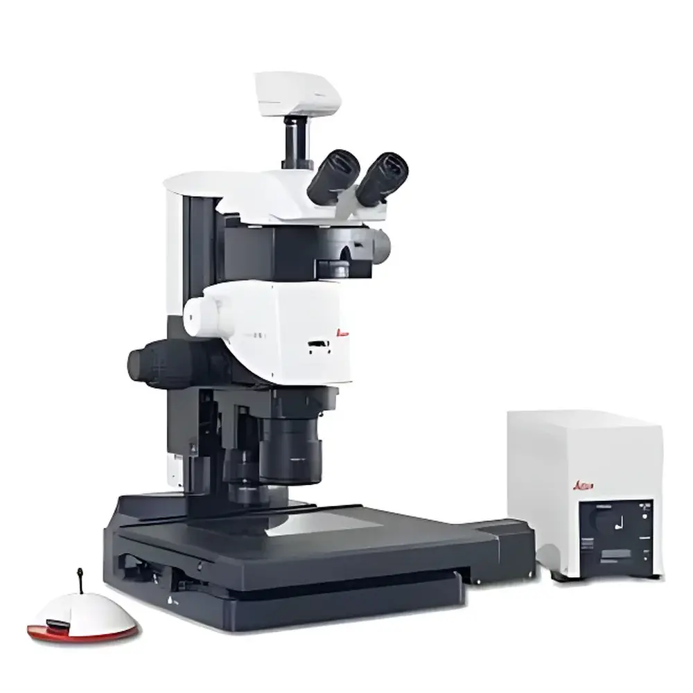
Leica M165 FC Fluorescence Stereo Microscope
| Brand | Leica |
|---|---|
| Origin | Singapore |
| Manufacturer Type | Authorized Distributor |
| Origin Category | Imported Instrument |
| Model | M165 FC |
| Price Range | USD 13,500 – 40,500 (FOB) |
| Instrument Type | Fluorescence Stereo Microscope |
| Excitation Source | High-Power LED-Based Fluorescence Illumination System (Compatible with Standard Filter Sets for DAPI, FITC, TRITC, Cy5) |
| Medical Device Classification | Not a Medical Device |
| Instrument Class | Research-Grade Fluorescence Stereo Microscope |
| Optical Zoom Ratio | 16.5:1 |
| Minimum Resolvable Feature Size | 1.1 µm (at maximum magnification with Plan Apo 2.0x objective and 10x eyepieces) |
| Image Encoding | Motorized, Encoder-Integrated Zoom & Focus Drive |
| Illumination Architecture | TripleBeam™ Optical Path Separation Technology |
| Imaging Mode Compatibility | Simultaneous Stereo Observation and Parallax-Free Digital Capture via FluoCombi™ Configuration |
| Modularity | Fully Modular Stand, Illumination, Camera, and Ergonomic Accessory Platform |
Overview
The Leica M165 FC is a research-grade fluorescence stereo microscope engineered for high-fidelity in vivo and ex vivo imaging of biological specimens requiring both three-dimensional spatial context and sensitive multi-channel fluorescence detection. Built upon Leica’s proprietary TripleBeam™ optical architecture, the system separates excitation light, observation light, and emission light into dedicated, non-interfering pathways—eliminating crosstalk, minimizing background noise, and maximizing signal-to-noise ratio across all fluorescence channels. Its apochromatic 16.5:1 zoom optics deliver diffraction-limited resolution down to 1.1 µm at full magnification (with Plan Apo 2.0× objective and 10× widefield eyepieces), enabling precise visualization of subcellular structures in intact tissues, zebrafish embryos, Drosophila larvae, plant meristems, and small-mammal surgical models. Unlike conventional stereo microscopes with shared illumination paths, the M165 FC maintains chromatic fidelity and geometric stability across the entire zoom range—critical for quantitative fluorescence intensity measurements and longitudinal time-lapse studies.
Key Features
- TripleBeam™ Illumination Architecture: Dedicated excitation path ensures uniform, collimated LED-based fluorescence illumination independent of observation optics—reducing stray light, improving contrast by >40% compared to dual-path systems, and enabling stable intensity calibration per channel.
- Apochromatic 16.5:1 Zoom Optics: Fully corrected for chromatic and spherical aberrations across visible and near-UV spectra; supports seamless transition from low-magnification specimen navigation (0.7×) to high-resolution structural analysis (11.5×).
- Motorized Image Encoding: Integrated position encoders on zoom, focus, and objective turrets automatically log magnification, working distance, illumination intensity, and filter configuration with every image—ensuring full metadata traceability compliant with GLP and ISO/IEC 17025 documentation requirements.
- FluoCombi™ Dual-Mode Imaging: Simultaneous binocular stereo viewing and parallax-free digital acquisition via coaxial camera port; eliminates registration errors during microinjection, electrophysiology, or laser ablation procedures.
- Modular Platform Design: Interchangeable stands (incl. vibration-damped, inclined, or inverted configurations), motorized stage options (XYZ translation, rotation, tilt), and accessory ports for transmitted light bases, polarization kits, and third-party environmental chambers.
Sample Compatibility & Compliance
The M165 FC accommodates live and fixed specimens ranging from whole-mount vertebrate embryos (mouse, chick, zebrafish) to dissected insect tissues, organoids, and 3D cell cultures. Its long working distance (up to 80 mm at 0.7×) and ergonomic sample access support micromanipulation workflows under sterile or hypoxic conditions. The system complies with IEC 61000-6-3 (EMC emissions), IEC 61000-6-2 (immunity), and EN 60601-1-2 (for lab-use safety). While not classified as a medical device per FDA 21 CFR Part 809 or EU IVDR, its hardware and firmware architecture meet foundational requirements for audit readiness in regulated preclinical research environments—including electronic record integrity (per ALCOA+ principles) and instrument configuration control.
Software & Data Management
Controlled via Leica Application Suite X (LAS X) Core or LAS X Live, the M165 FC supports automated multichannel acquisition, Z-stack generation, extended depth-of-field (EDF) reconstruction, and real-time spectral unmixing when paired with appropriate sCMOS or EMCCD cameras. All image metadata—including encoder-derived magnification, exposure time, LED power level, filter wheel position, and stage coordinates—is embedded in TIFF and OME-TIFF formats. LAS X includes built-in tools for batch processing, ROI-based intensity quantification, and export to HDF5 for integration with Python- or MATLAB-based analysis pipelines. Audit trail functionality logs user actions, parameter changes, and system events—fully compatible with 21 CFR Part 11-compliant electronic signature deployment when configured with validated server infrastructure.
Applications
- Developmental biology: Time-lapse imaging of fluorescent reporter expression in live embryos
- Neuroscience: Stereotactic injection guidance and post-hoc verification of AAV transduction patterns
- Oncology research: Visualizing tumor xenograft vascularization using lectin or CD31 immunofluorescence
- Plant science: Root architecture phenotyping with GFP-tagged auxin transporters
- Regenerative medicine: Monitoring stem-cell-derived organoid maturation and lumen formation
- Quality control in bioproduction: Rapid visual assessment of cell pellet integrity and contamination screening
FAQ
Is the M165 FC suitable for quantitative fluorescence intensity measurements?
Yes—its encoded zoom/focus system, calibrated LED excitation output, and consistent optical throughput across magnifications enable reproducible relative intensity comparisons within and between experiments.
Can the system be integrated with third-party environmental control chambers?
Yes—the modular stand design includes standardized mounting interfaces (M6 and M4 threaded holes) and cable routing channels compatible with commercial incubation, CO₂, and temperature-control enclosures.
Does Leica provide validation documentation for GxP environments?
Leica offers IQ/OQ protocols and URS templates upon request; full PQ must be performed in situ by the end user or qualified service provider per local regulatory expectations.
What camera interfaces are supported?
USB 3.0, Camera Link, and GigE Vision—compatible with scientific CMOS, EMCCD, and sCMOS sensors from Hamamatsu, Photometrics, and Andor.
Is motorized focus included as standard equipment?
Motorized focus is optional but highly recommended for encoded workflows; it is required for full LAS X automation and EDF acquisition.


