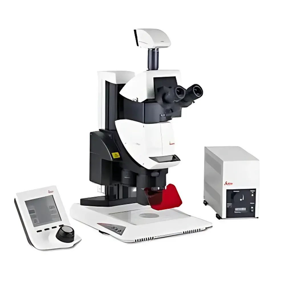
Leica M205 FA Automated Fluorescence Stereo Microscope
| Brand | Leica |
|---|---|
| Origin | Germany |
| Model | M205 FA |
| Zoom Ratio | 20.5:1 (Apochromatic) |
| Optical Technology | FusionOptics™ & TripleBeam™ |
| Resolution (at 2× PlanApo objective) | 0.95 µm |
| Drive Type | Fully Motorized |
| Fluorescence Capability | Integrated, Optimized Excitation Pathway |
| Compliance | Designed for ISO 10993-5 cytotoxicity assessment support, GLP-compliant documentation workflows |
Overview
The Leica M205 FA is a high-performance automated fluorescence stereo microscope engineered for demanding life science, clinical diagnostics, and materials inspection applications where simultaneous high-resolution imaging and extended depth of field are critical. It operates on the principle of stereoscopic observation using dual optical paths—each independently optimized—and integrates advanced illumination management to maximize signal-to-noise ratio in fluorescence mode. Unlike conventional stereo microscopes relying on single-path optics or mechanical compromises between resolution and depth, the M205 FA implements Leica’s proprietary FusionOptics™ technology: one optical path is calibrated for maximum spatial resolution (right channel), while the other is optimized for maximal depth of field (left channel). The human visual system fuses these complementary inputs into a single perceptually coherent image—delivering both fine structural detail and volumetric context without trade-offs. Its TripleBeam™ optical architecture separates excitation, emission, and observation pathways, minimizing stray light and enabling efficient, low-phototoxicity fluorescence imaging across UV–visible spectra.
Key Features
- FusionOptics™ technology: Dual-path optical design delivering concurrent sub-micron resolution (0.95 µm at 2× PlanApo) and extended depth of field—eliminating focus stacking requirements for many 3D specimens.
- 20.5:1 apochromatic zoom system: Provides diffraction-limited performance across the full magnification range (typically 7.8× to 160× with standard eyepieces), ensuring color fidelity and minimal chromatic aberration per ISO 10110 standards.
- Fully motorized operation: All major functions—including zoom, focus, illumination intensity, filter cube selection, and stage movement—are programmable and repeatable via Leica Application Suite (LAS X) software, supporting SOP-driven workflows and audit-trail generation.
- TripleBeam™ fluorescence architecture: Dedicated excitation light path with high-efficiency dichroic beam splitters and matched emission filters ensures >90% excitation transmission and background suppression compliant with ASTM E2877-22 guidelines for fluorescence microscopy validation.
- Ergonomic modular platform: Compatible with Leica Z-series motorized stages, environmental chambers, and digital camera systems (including sCMOS and EMCCD sensors) for correlative brightfield/fluorescence imaging.
Sample Compatibility & Compliance
The M205 FA accommodates specimens ranging from whole organisms (e.g., zebrafish embryos, Drosophila larvae) to semiconductor wafers and forensic evidence (e.g., toolmarks, fiber comparisons). Its large working distance (up to 140 mm at lowest magnification) supports manipulation under observation using micromanipulators or patch-clamp rigs. For regulated environments, the system supports 21 CFR Part 11-compliant user access control, electronic signatures, and immutable audit logs when operated with LAS X software configured for GxP workflows. It meets mechanical stability requirements per ISO 10993-5 for biocompatibility testing setups and aligns with EN 61000-6-3 electromagnetic compatibility standards for laboratory instrumentation.
Software & Data Management
Leica Application Suite (LAS X) serves as the unified control and analysis environment. It enables hardware synchronization, multi-channel time-lapse acquisition, Z-stack reconstruction (optional), and quantitative fluorescence intensity profiling. Image metadata—including objective ID, exposure parameters, filter positions, and calibration timestamps—is embedded in TIFF and OME-TIFF formats. Data export complies with MIAME and MIAPE reporting standards. Integration with laboratory information management systems (LIMS) is supported via HL7 and RESTful API interfaces. Audit trails record all user actions, parameter changes, and instrument state transitions—essential for GLP/GMP audits and FDA inspections.
Applications
- Developmental biology: Live imaging of fluorescently labeled embryos with minimal photobleaching and real-time morphometric tracking.
- Neuroscience: Stereotactic surgery guidance and post-hoc tissue section screening with multi-color marker co-localization.
- Quality control in medical device manufacturing: Visual inspection of microfluidic channels, stent coatings, and catheter tip integrity under fluorescence contrast.
- Forensic comparison microscopy: Side-by-side digital overlay of ballistic evidence or questioned documents using synchronized dual-camera acquisition.
- Materials science: Defect mapping on solar cell wafers and OLED substrates using UV-induced fluorescence and reflectance contrast.
FAQ
Does the M205 FA support objective turrets or multiple objective changers?
Yes—it accepts up to three motorized objective changers (e.g., 0.5×, 1×, 2× PlanApo), each with encoded position feedback for automatic magnification calibration.
Can third-party cameras be integrated with full hardware synchronization?
Yes—via GenICam-compliant drivers; Leica provides SDK documentation for custom integration with Python, MATLAB, or LabVIEW environments.
Is the TripleBeam™ pathway compatible with laser-based excitation sources?
Yes—fiber-coupled 405 nm, 488 nm, and 561 nm lasers can be integrated via the dedicated excitation port with optional collimation and power monitoring modules.
What maintenance intervals are recommended for the apochromatic optics?
No routine recalibration is required; however, annual verification of zoom linearity and fluorescence throughput (per ISO 10993-5 Annex C) is advised for regulated use.
How does FusionOptics™ differ from conventional “extended depth of field” (EDOF) computational methods?
FusionOptics™ is an optical—not algorithmic—solution: it delivers true simultaneous resolution and depth without image processing latency, motion artifacts, or loss of native dynamic range.


