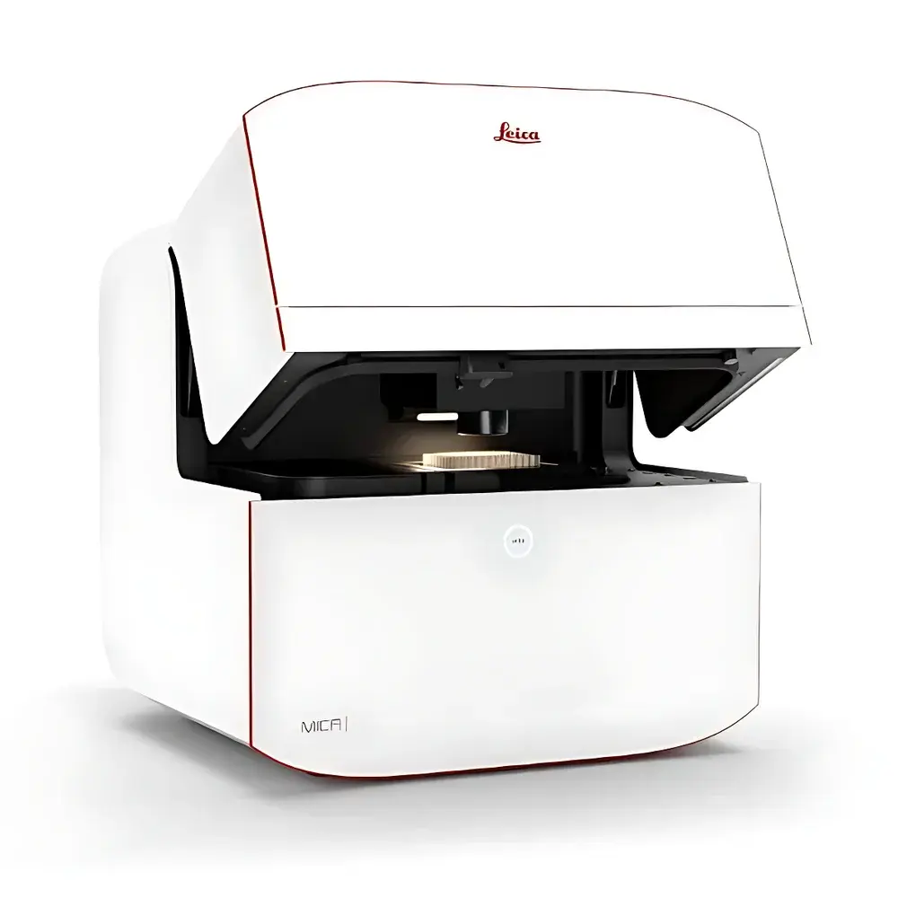
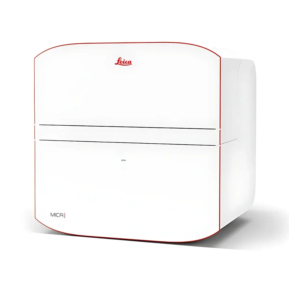
Leica MICA Widefield Multimodal Imaging Analysis Platform
| Brand | Leica |
|---|---|
| Origin | Germany |
| Manufacturer Type | Authorized Distributor |
| Product Category | Imported Instrument |
| Model | MICA Widefield |
| Instrument Type | Inverted Fluorescence Microscope |
| Excitation Source | High-Power LED |
| Objective Options | 63× Water Immersion Objective (CS2, NA 1.20) with Auto-Water Delivery |
| Fluorescence Detection Technology | FluoSync Hyperspectral Unmixing |
| Control Architecture | Fully Motorized & Software-Driven |
| Focusing Strategy | Three Adaptive Focus Modes (Overview, Detail, Subcellular) |
| Observation Interface | Integrated Scientific CMOS Camera |
| DIC Compatibility | Optimized for Glass and Plastic Culture Vessels |
| Stage | Fully Motorized XY Scanning Stage |
| Filter System | FluoSync-Enabled Spectral Separation Module |
Overview
The Leica MICA Widefield Multimodal Imaging Analysis Platform represents a paradigm shift in biological microscopy—redefining accessibility, reproducibility, and analytical rigor for both novice and expert users. Engineered as a unified imaging analysis hub, MICA integrates widefield fluorescence, confocal point-scanning, transmitted light (DIC/Phase), and intelligent computational imaging into a single, cohesive system. Its core architecture leverages optical coherence across modalities to ensure absolute spatiotemporal correlation of multichannel data—eliminating registration artifacts inherent in sequential acquisition. Unlike conventional inverted microscopes requiring manual alignment, calibration, and iterative parameter tuning, MICA embeds hardware-software co-optimization at every layer: from LED excitation intensity and spectral unmixing to adaptive focus control and environmental regulation. Designed for live-cell and fixed-tissue applications alike, it operates within a fully enclosed incubation chamber enabling precise regulation of temperature (±0.2 °C), CO₂ (0–20%), and humidity—critical for long-term time-lapse experiments exceeding 72 hours.
Key Features
- Fully Motorized & AI-Guided Workflow: All optical components—including objective turret, filter cubes, focus drive, stage, and illumination—are motorized and synchronized via Leica’s proprietary software platform. OneTouch automation dynamically adjusts exposure, gain, LED power, and z-stack parameters based on user-defined priorities (e.g., “Sample Protection” vs. “Image Quality”).
- FluoSync Hyperspectral Unmixing: A patented hardware-software hybrid approach enabling simultaneous detection and real-time spectral decomposition of up to four fluorophores without temporal or spatial offset. Combines a tunable emission filter wheel with advanced linear unmixing algorithms trained on reference spectra.
- Adaptive Focus Strategies: Three context-aware focusing modes—“Overview”, “Detail”, and “Subcellular”—automatically adjust step size, contrast threshold, and search range to maintain optimal focus across heterogeneous samples and time-lapse series.
- Integrated Live-Cell Incubation Chamber: Sealed environmental control unit maintains physiological conditions during extended acquisitions. Includes vapor-trap design reducing medium evaporation by >60% over standard open-stage systems.
- THUNDER & LIGHTNING Computational Imaging: THUNDER deconvolution enhances optical sectioning in widefield mode; LIGHTNING delivers sub-diffraction detail reconstruction in confocal mode—all embedded natively within the acquisition pipeline without post-hoc processing delays.
- Pixel Classifier AI Training Interface: Enables supervised machine learning directly within the GUI. Users annotate regions-of-interest using intuitive drawing tools; the system trains a reusable pixel classifier model compliant with FAIR data principles and GLP audit trails.
Sample Compatibility & Compliance
MICA supports a broad range of specimen formats including glass-bottom dishes, multi-well plates (6–384-well), organ-on-chip devices, and thick tissue sections (up to 200 µm). Its DIC-compatible optics accommodate both glass and polymer-based culture substrates without compromise in contrast or resolution. The platform conforms to ISO 13485:2016 for medical device quality management and supports FDA 21 CFR Part 11-compliant electronic records through encrypted audit logs, user authentication, and immutable acquisition metadata embedding. All image data is stored in OME-TIFF format, ensuring interoperability with OMERO, QuPath, and other open-source analysis ecosystems.
Software & Data Management
Acquisition, analysis, and reporting are unified under Leica Application Suite X (LAS X) v4.13+, featuring native integration of AI-assisted segmentation, batch processing pipelines, and export-ready report generation. Raw data—including full spectral stacks, z-series, and time-lapse metadata—is automatically archived with SHA-256 checksums and timestamped provenance tracking. The system supports DICOM-SR export for clinical research applications and provides RESTful API access for LIMS integration. All AI models generated via Pixel Classifier are version-controlled, exportable as .zip packages, and validated per ISO/IEC 17025 guidelines for method verification.
Applications
- High-Content Screening (HCS): Simultaneous four-channel acquisition in multi-well plates enables kinetic assays such as caspase-3/7 activation, mitochondrial membrane potential (TMRE), cytoskeletal dynamics (SiR-Actin), and nuclear morphology (DAPI)—all with zero temporal drift.
- 3D Tissue Imaging: Seamless transition from low-magnification overview (20×) to high-resolution confocal (63× water) allows hierarchical interrogation of intestinal crypt architecture, tubulin detyrosination patterns, and cell-type-specific marker colocalization.
- Long-Term Live-Cell Dynamics: Sustained GFP expression monitoring in MDCK spheroids over 72+ hours demonstrates minimal phototoxicity and stable environmental maintenance—validated against ASTM E3085-17 standards for bioreactor mimicry.
- Multiplexed Subcellular Phenotyping: Combined use of THUNDER (for volumetric reconstruction) and LIGHTNING (for ultrastructural enhancement) enables quantitative assessment of organelle morphology, vesicle trafficking, and protein aggregation kinetics.
FAQ
What distinguishes FluoSync from conventional spectral unmixing methods?
FluoSync couples physical bandpass filtering with real-time constrained linear unmixing, eliminating the need for separate reference scans and reducing acquisition time by up to 4× compared to sequential filter-based approaches.
Can MICA be used for GMP-regulated workflows?
Yes—when deployed with optional Leica Validation Package, MICA meets requirements for instrument qualification (IQ/OQ/PQ), electronic signature enforcement, and 21 CFR Part 11 compliance.
Is the 63× water immersion objective compatible with automated water replenishment during time-lapse?
Yes—the CS2 objective features integrated fluidics that maintain meniscus stability across multi-hour acquisitions without manual intervention.
How does MICA ensure reproducibility across users and laboratories?
Through standardized acquisition templates, versioned AI models, and embedded metadata capturing all hardware states, environmental parameters, and software settings—enabling full experimental traceability per ISO/IEC 17025 clause 7.7.
Does MICA support third-party camera or detector integration?
No—MICA uses a purpose-built sCMOS sensor optimized for FluoSync spectral response and dynamic range; external detectors are not supported to preserve calibration integrity and regulatory compliance.


