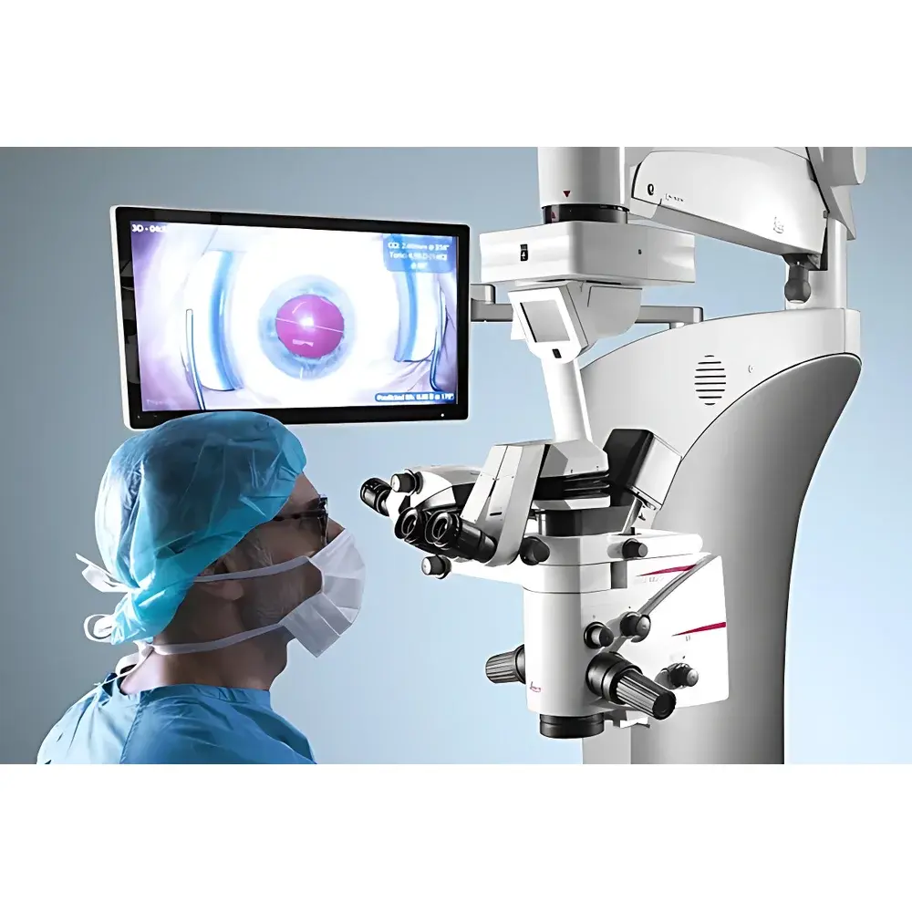
Leica Proveo 8 Ophthalmic Surgical Microscope
| Brand | Leica |
|---|---|
| Origin | Germany |
| Model | Proveo 8 |
| Application | Ophthalmic Surgery |
| Optical Architecture | CoAx4 Illumination & FusionOptics Dual-Path System |
| Integrated Imaging | Optional iOCT (Intraoperative Optical Coherence Tomography) |
| Ergonomic Design | Extended Arm Reach (+21%), Reduced Base Footprint (−33%) |
| User Interface | Touchscreen Control Panel & Footswitch-Activated Workflow Presets |
| Compliance | CE Marked, ISO 13485 Certified, Designed for GLP/GMP-Aligned Clinical Environments |
Overview
The Leica Proveo 8 Ophthalmic Surgical Microscope is an advanced, modular surgical visualization platform engineered specifically for anterior and posterior segment ophthalmic procedures. It operates on dual optical principles: CoAx4 — a four-channel coaxial LED illumination architecture delivering uniform, glare-free red reflex across the entire surgical field — and FusionOptics, a patented dual-light-path system that simultaneously optimizes depth of field (+40%) and spatial resolution in real time. Unlike conventional binocular microscopes relying on single-path optics with inherent trade-offs between focus depth and detail clarity, the Proveo 8 synthesizes two independently optimized image streams into a single stereoscopic percept, enabling surgeons to maintain precise focus from scleral entry through vitreoretinal interface without manual refocusing. This architecture directly addresses the clinical challenge of low-light, high-precision interventions where visual fidelity correlates with procedural accuracy, intraoperative decision latency, and tissue preservation.
Key Features
- CoAx4 Illumination: Four independent coaxial LED channels deliver consistent red reflex throughout cataract surgery—including phacoemulsification—eliminating shadowing, minimizing phototoxicity risk, and preserving natural tissue contrast under reduced luminance settings.
- FusionOptics Technology: Separates optical pathways for depth perception and fine-detail resolution; one path extends focal volume while the other maximizes resolving power at the target plane—automatically fused by the visual cortex into a unified 3D representation.
- Modular Integration Framework: Supports seamless docking of intraoperative imaging modalities including Leica’s iOCT system for real-time cross-sectional visualization of corneal layers, capsular bag dynamics, and retinal morphology during live surgery.
- Ergonomic Redesign: Increased arm reach (21% longer than prior-generation platforms) and 33% smaller base footprint improve operating room spatial flexibility and reduce collision risk with adjacent equipment or personnel.
- Workflow-Centric Controls: Programmable footswitch presets allow rapid transition between pre-configured surgical stages (e.g., capsulorhexis → hydrodissection → phaco → IOL insertion); all parameters displayed on integrated touchscreen status panel for immediate verification.
Sample Compatibility & Compliance
The Proveo 8 is validated for use across full-spectrum ophthalmic interventions: anterior segment procedures (cataract extraction, corneal transplantation, gonioscopy-assisted surgery), vitreoretinal surgery (membrane peeling, endolaser application, macular hole repair), and combined anterior-posterior approaches. Its optical calibration adheres to DIN EN ISO 10940 (Ophthalmic instruments — Requirements for surgical microscopes) and conforms to IEC 60601-2-46 for medical electrical equipment safety. The system supports audit-trail-enabled documentation workflows compliant with FDA 21 CFR Part 11 requirements when paired with Leica’s certified DICOM-compliant image archiving modules. All illumination intensity settings are traceable and reproducible per ANSI Z80.10 standards for ophthalmic diagnostic equipment.
Software & Data Management
Embedded firmware enables synchronized capture of high-fidelity 4K video, stereo stills, and optional OCT cross-sections via Leica’s LAS X software suite. Image metadata—including magnification level, illumination intensity, zoom position, and timestamped workflow stage—is automatically embedded and exportable in DICOM-SR format. Remote collaboration functionality permits encrypted streaming to PACS networks or tele-mentoring platforms meeting HIPAA and GDPR data handling protocols. Software updates are delivered through secure, version-controlled firmware packages with rollback capability—ensuring continuity in regulated clinical environments.
Applications
- Cataract surgery with real-time assessment of capsulorhexis integrity and lens fragment dynamics
- Vitrectomy with enhanced visualization of epiretinal membranes, internal limiting membrane, and subretinal hemorrhage margins
- Endothelial keratoplasty (DSAEK/DMEK) requiring micron-level graft orientation and apposition confirmation
- Glaucoma drainage device implantation under direct visualization of Schlemm’s canal anatomy
- Intraoperative teaching scenarios leveraging heads-up 3D display integration for trainee engagement without compromising primary surgeon ergonomics
FAQ
Is the Proveo 8 compatible with existing Leica surgical accessories and footswitches?
Yes—the platform maintains mechanical and electrical backward compatibility with Leica’s universal mounting interfaces and legacy control peripherals, subject to firmware version alignment.
Does FusionOptics require special training for effective utilization?
No—FusionOptics operates transparently at the optical level; no user calibration or adjustment is needed. Surgeons perceive improved stereopsis and extended depth-of-field intuitively, without altering standard surgical technique.
Can iOCT be retrofitted to an installed Proveo 8 unit?
Yes—iOCT integration is available as a field-upgradeable module, provided the microscope’s optical bench and control unit meet minimum revision thresholds specified in Leica Service Bulletin L-PROV-2023-07.
How does CoAx4 illumination impact photobiomodulation safety during prolonged procedures?
CoAx4 utilizes narrow-band red LEDs (625 ± 5 nm) with peak irradiance below ICNIRP 2013 exposure limits for retinal photic injury; total intraocular radiant exposure remains within Class 1 laser safety classification per IEC 60825-1.
What documentation support is available for regulatory submissions in FDA-regulated markets?
Leica provides comprehensive technical files, design history documentation (DHF), and risk management reports aligned with ISO 14971:2019, supporting 510(k) or De Novo pathway submissions for clinical deployment in U.S. healthcare institutions.
Related Products


