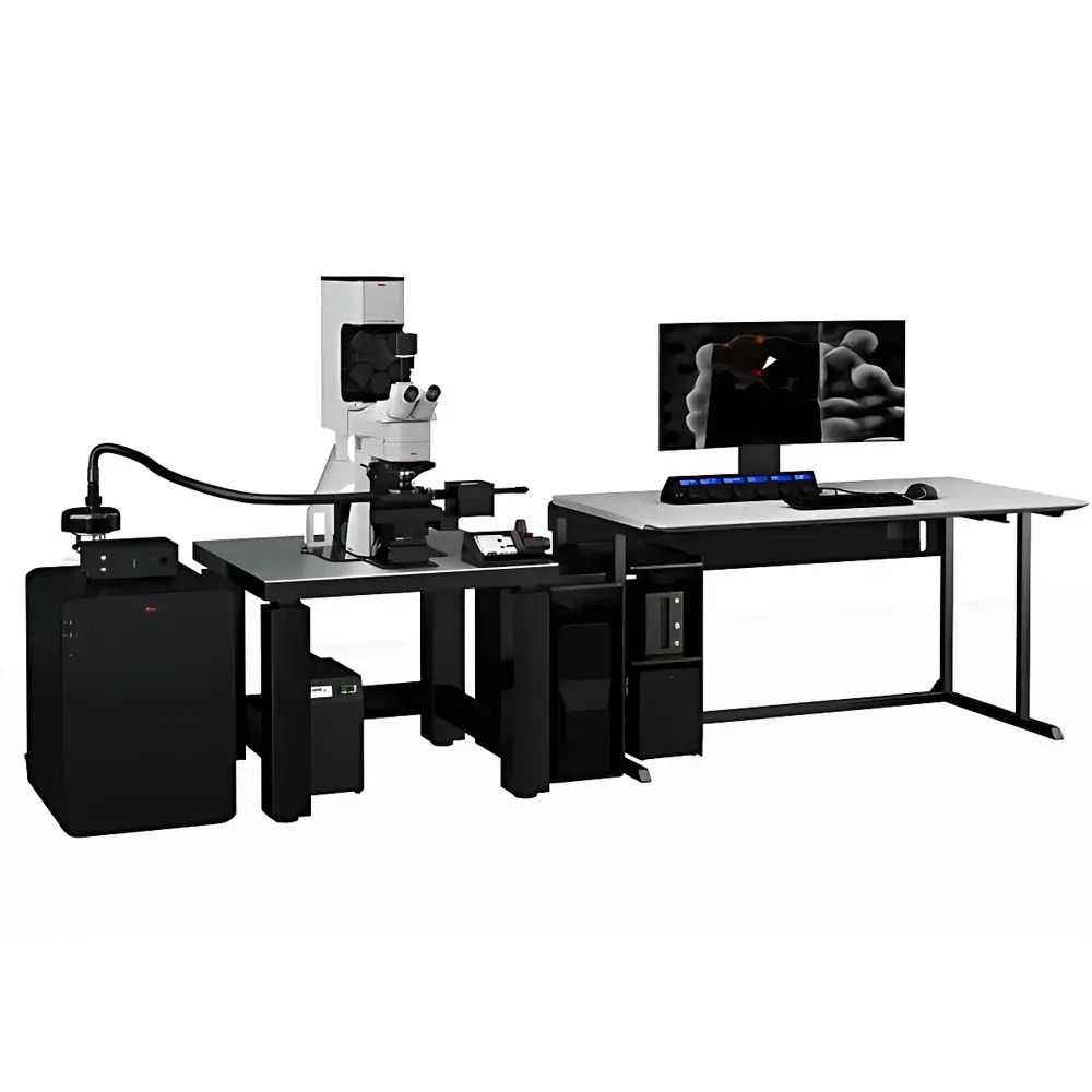
Leica STELLARIS Cryo Confocal Microscope
| Brand | Leica |
|---|---|
| Origin | Germany |
| Model | STELLARIS Cryo |
| Application Domain | Cryo-Correlative Light and Electron Microscopy (Cryo-CLEM) |
| Cooling Stage | Integrated Liquid Nitrogen–Cooled Stage with Transfer Shuttle |
| Software Platform | LAS X Coral Cryo (with TauSense, LIGHTNING, and Interpolated 3D Targeting) |
| Objective Lens | HC PL APO 50×/0.90 Cryo-Optimized Dry Objective |
| Detection System | Power HyD Hybrid Detectors |
| Light Source | White Light Laser (WLL, 470–670 nm) |
| Imaging Modes | Confocal Reflectance, Fluorescence, TauContrast, TauSeparation, Spectral Unmixing |
| Ice Thickness Assessment | Dual-mode (Overview + Confocal Reflectance Z-Stack) |
| Data Format | Open, Metadata-Rich TIFF & HDF5 (CF-1 compliant) |
| Regulatory Alignment | Designed for GLP/GMP-aligned workflows |
Overview
The Leica STELLARIS Cryo Confocal Microscope is a purpose-built optical imaging platform engineered for cryo-correlative light and electron microscopy (Cryo-CLEM), specifically to enable high-precision, three-dimensional target localization in vitrified biological specimens prior to cryo-electron tomography (CryoET). Unlike conventional confocal systems, the STELLARIS Cryo integrates a fully enclosed, liquid nitrogen–cooled stage and transfer shuttle to maintain samples continuously below −160 °C throughout optical interrogation, sample navigation, and coordinate registration. Its core measurement principle relies on diffraction-limited laser scanning fluorescence combined with confocal reflectance imaging—both performed under cryogenic conditions—to generate spatially registered, open-format coordinate maps (x, y, z, θ) that are directly translatable into focused ion beam (FIB) milling or transmission electron microscopy (TEM) grid navigation protocols. This ensures deterministic retrieval of fluorescently labeled structures within the same physical volume later imaged by electrons—where fluorescence signals are no longer detectable.
Key Features
- Integrated cryogenic stage with active liquid nitrogen cooling (≤ −180 °C base temperature) and inert gas overpressure environment to prevent frost formation during stage movement and objective focusing
- Cryo-optimized HC PL APO 50×/0.90 dry objective with extended working distance and minimized thermal drift—designed for high-resolution imaging without immersion media or condensation risk
- White Light Laser (WLL) excitation source (470–670 nm) enabling flexible, multi-color fluorophore excitation while minimizing photodamage in frozen-hydrated samples
- Power HyD hybrid detectors with single-photon sensitivity and time-resolved capability (TCSPC-ready), supporting both intensity-based and lifetime-based contrast generation
- TauSense technology: simultaneous acquisition of spectral emission profiles and photon arrival time distributions (τ), enabling fluorophore unmixing via TauContrast and TauSeparation—even when spectral overlap occurs due to cryo-induced spectral shifts
- LAS X Coral Cryo software suite featuring interpolated 3D targeting: reconstructs precise fiducial coordinates from multi-layer z-stacks using sub-pixel interpolation algorithms, outputting CF-1–compliant coordinate files for downstream FIB-SEM or TEM stage control
- Dual-mode ice thickness assessment: rapid widefield overview mode for gross ice quality screening, plus confocal reflectance z-stack analysis to quantify ice layer thickness (±5 nm resolution) in 3D prior to milling
Sample Compatibility & Compliance
The STELLARIS Cryo accommodates standard EM grids (e.g., Quantifoil R2/2, UltrAuFoil) mounted on custom cryo-shuttles compatible with Leica EM VCT100 or Thermo Fisher AutoSlice & View workflows. Sample handling conforms to ISO 13843:2021 (cryomicroscopy terminology and metrology) and supports traceable alignment to EM coordinate systems via calibrated stage encoders (resolution ≤ 50 nm). All metadata—including temperature logs, laser power settings, detector gain, and stage position timestamps—are embedded in HDF5 and TIFF outputs per the Common Format for Cryo-EM (CF-1) specification. When configured with validated LAS X Coral Cryo installation and electronic signature modules, the system meets documentation requirements for GLP-compliant structural biology studies and aligns with FDA 21 CFR Part 11 principles for audit-trail integrity in regulated environments.
Software & Data Management
LAS X Coral Cryo provides a unified interface for experimental design, acquisition, and coordinate export. Its modular architecture includes dedicated tools for cryo-specific QA: ice-thickness heatmaps, fiducial signal-to-noise ratio (SNR) scoring, and automatic drift correction across multi-hour acquisitions. All image data are saved in open, self-describing formats (TIFF + sidecar JSON / HDF5), ensuring interoperability with third-party reconstruction pipelines (e.g., IMOD, Dynamo, pySEMS). The software supports batch processing of z-stacks for TauContrast quantification and exports coordinate lists in .csv and .xml formats compatible with Thermo Fisher Maps, Zeiss Atlas 5, and Delmic SECOM platforms. Optional integration with laboratory information management systems (LIMS) enables automated metadata ingestion and workflow tracking.
Applications
- Precise targeting of subcellular organelles (e.g., mitochondria, endosomes, synaptic vesicles) for site-specific CryoET lamella preparation
- Validation of cryo-FIB milling depth and orientation relative to fluorescent landmarks
- Quantitative assessment of ice contamination and vitrification quality across grid squares
- Multi-parametric phenotyping of frozen cellular states using lifetime-resolved contrast (e.g., distinguishing protein conformational states via τ-shifted fluorophores)
- Correlative validation of CRISPR/Cas9 editing outcomes in primary neurons or organoids at near-native structural preservation
- Time-resolved Cryo-CLEM of dynamic processes arrested by plunge-freezing (e.g., clathrin-coated pit assembly, viral entry intermediates)
FAQ
What distinguishes STELLARIS Cryo from standard confocal microscopes?
It features a fully integrated, vibration-isolated cryostage, cryo-optimized optics, and software-engineered coordinate export protocols—none of which are present in ambient-temperature confocal platforms.
Can STELLARIS Cryo be used without a cryo-EM facility?
Yes—it functions as a standalone cryo-fluorescence microscope for ice quality assessment, fiducial mapping, and live–frozen correlation studies, though its full value is realized in integrated Cryo-CLEM workflows.
Does LAS X Coral Cryo support automated acquisition scripts?
Yes—Python-based scripting via the LAS X API allows programmable grid navigation, multi-position z-stack acquisition, and conditional triggering based on real-time SNR or ice-thickness thresholds.
How is coordinate accuracy validated?
Through independent calibration using gold nanoparticle fiducials on EM grids, cross-verified against post-milling TEM images; typical reproducibility is ≤ 100 nm across repeated acquisitions under identical thermal conditions.
Is TauSense data acquisition compatible with low-light cryo conditions?
Yes—Power HyD detectors operate at >30% quantum efficiency down to single-photon flux levels, and TauSense processing uses photon arrival time histograms accumulated over defined dwell times—not instantaneous frame rates—ensuring robustness under cryo photon starvation constraints.


