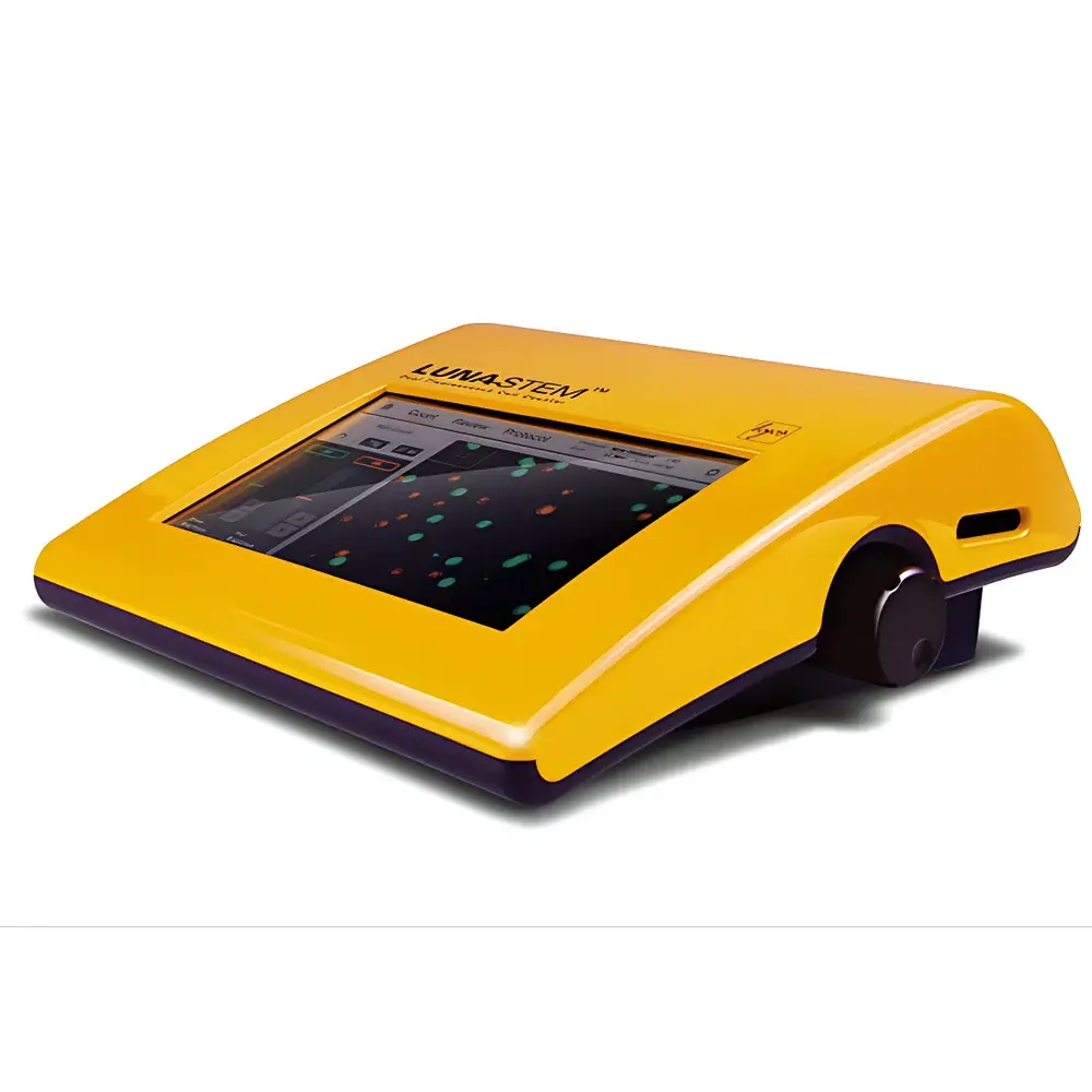
LUNA-STEM™ L30001 Automated Fluorescence Cell Counter
| Brand | Aligned Genetics |
|---|---|
| Origin | South Korea |
| Model | L30001 |
| Detection Time | 30–60 s |
| Cell Concentration Range | 5 × 10⁴ – 1 × 10⁷ cells/mL |
| Sample Volume | 10 µL |
| Detectable Cell Size Range | 1–90 µm |
| Excitation Wavelength | 470 ± 20 nm |
| Emission Wavelengths | 530 ± 25 nm and 600 nm (long-pass) |
| Light Source | LED |
| Image Resolution | 5 MP |
| Display | 800 × 480 pixels |
| Dimensions (W×D×H) | 22 × 21 × 9 cm |
| Weight | 1.8 kg |
Overview
The LUNA-STEM™ L30001 Automated Fluorescence Cell Counter is an engineered solution for precise, rapid, and reproducible quantification of heterogeneous cell populations—particularly adipose-derived stem cells (ADSCs) and stromal vascular fraction (SVF) isolates. It operates on a dual-channel fluorescence + brightfield imaging principle: nucleated cells are identified and differentiated by membrane-impermeant and membrane-permeant nucleic acid dyes, while enucleated particles (e.g., platelets, erythrocyte ghosts, or debris) are resolved via morphological and optical contrast in brightfield. This tri-modal acquisition (green fluorescence for live nucleated cells, red fluorescence for dead nucleated cells, and brightfield for total particulate objects) enables simultaneous enumeration of viable nucleated cells, non-viable nucleated cells, and enucleated events—all within a single 10 µL sample load. The system is optimized for primary tissue-derived suspensions where conventional hemocytometry or single-channel counters fail to resolve overlapping populations with high fidelity.
Key Features
- Dual-fluorescence + brightfield optical architecture ensures unambiguous discrimination between live nucleated cells, dead nucleated cells, and enucleated particles based on dye exclusion, nuclear morphology, and size distribution.
- Integrated 5-megapixel CMOS imaging sensor captures high-fidelity, focus-stabilized images across all three channels without user intervention.
- Automated image registration and pixel-level channel fusion enables robust segmentation of low-contrast or clustered cells common in SVF preparations.
- Onboard analysis engine computes total cell concentration, nucleated cell concentration, enucleated particle concentration, viability percentage, and mean cell diameter—all reported in ≤60 seconds.
- Self-contained operation with built-in 800 × 480 LCD touchscreen interface; no external computer required for routine counting or report generation.
- LED-based excitation source (470 ± 20 nm) and dual-band emission detection (530 ± 25 nm and 600 nm long-pass) ensure stable, low-heat illumination with minimal photobleaching during repeated assays.
Sample Compatibility & Compliance
The LUNA-STEM™ L30001 is validated for use with enzymatically digested adipose tissue isolates, bone marrow mononuclear cells (MNCs), umbilical cord blood derivatives, and other primary suspension samples containing mixed nucleated/enucleated populations. It accommodates cells ranging from 1 µm (small platelet fragments) to 90 µm (large adipocyte-derived microvesicles or aggregated stromal cells), with optimal quantitative accuracy between 5–60 µm. The instrument complies with IEC 61010-1 for laboratory equipment safety and meets electromagnetic compatibility (EMC) requirements per EN 61326-1. While not certified as a medical device under FDA 21 CFR Part 820 or ISO 13485, its data output structure supports GLP-compliant documentation when integrated into validated laboratory workflows. Audit-trail-capable PDF reports include timestamped raw images, metadata (sample ID, operator, date/time), and full parameter logs—facilitating traceability in regulated research environments.
Software & Data Management
All image capture, segmentation, and calculation routines are executed onboard the device firmware—eliminating dependency on proprietary desktop software or cloud services. Counting results and annotated images are exported as password-protected PDF reports directly to USB storage devices. Each report contains: (i) composite RGB-merged visualization of all three channels; (ii) scatterplots of area vs. fluorescence intensity for population gating validation; (iii) tabulated numerical outputs with confidence intervals derived from statistical sampling of ≥500 detected objects per field; and (iv) operator-entered sample annotations. Data export conforms to FAIR principles (Findable, Accessible, Interoperable, Reusable) through standardized metadata tagging (ISO/IEC 11179-compliant descriptors). No telemetry, remote access, or automatic data upload occurs—ensuring full local control over sensitive biological data.
Applications
- Quantitative release testing of SVF and ADSC products prior to expansion or transplantation.
- Monitoring culture health during early passage expansion—detecting spontaneous enucleation or fragmentation events.
- Standardizing input cell numbers for flow cytometry, RNA extraction, or functional assays requiring nucleated-cell-specific normalization.
- Quality control of cryopreserved samples—assessing post-thaw viability and particulate contamination.
- Method comparison studies against manual hemocytometry or single-dye automated counters (e.g., AO/PI-only platforms).
- Training and standardization across multi-site regenerative medicine laboratories seeking inter-operator consistency.
FAQ
What cell types are specifically validated for use with the LUNA-STEM™ L30001?
Adipose-derived stem cells (ADSCs), stromal vascular fraction (SVF) isolates, bone marrow mononuclear cells (MNCs), and umbilical cord blood-derived mononuclear preparations have been experimentally verified for accurate nucleated/enucleated discrimination.
Does the instrument require calibration with reference beads or control samples?
No routine calibration is required; the system employs factory-characterized optical path geometry and fixed LED intensity profiles. Optional daily verification using standardized polystyrene microsphere suspensions (10 µm and 25 µm) is supported for QC documentation.
Can the LUNA-STEM™ distinguish between apoptotic and necrotic cells?
It differentiates membrane-intact (live) versus membrane-compromised (dead) nucleated cells using standard viability dyes (e.g., acridine orange/propidium iodide or similar). Apoptosis-specific markers (e.g., Annexin V) require compatible secondary staining protocols but are not automatically classified by default algorithms.
Is the device compatible with Good Manufacturing Practice (GMP) environments?
While not GMP-certified, its design supports GMP-aligned practices: electronic records meet ALCOA+ criteria (Attributable, Legible, Contemporaneous, Original, Accurate, Complete, Consistent, Enduring, Available), and audit logs are immutable once generated.
What is the recommended maintenance schedule?
Optical surfaces should be cleaned weekly with lint-free wipes and isopropyl alcohol; LED lifetime exceeds 10,000 hours; no consumable parts require scheduled replacement within the first 36 months of typical lab use.


