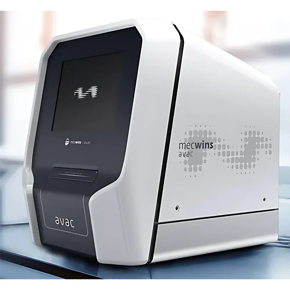
Mecwins AVAC High-Throughput Single-Molecule Immunoassay Analyzer
| Brand | Mecwins |
|---|---|
| Origin | Spain |
| Model | AVAC |
| Detection Principle | Plasmonic Nanoparticle Scattering Spectroscopy |
| Sensitivity | 3 fM LoD (miRNA-122) |
| Throughput | 96-well plate in 5 min |
| Max Image Acquisition Rate | 20,000 images/hour |
| Multiplex Capacity | Up to 5 analytes per sample |
| Counting Range | 10²–10⁶ molecules/field |
| Spatial Resolution | 0.7 µm (diffraction-limited) |
| Substrate | Functionalized polymer matrix (silicon-free) |
| Automation Level | Fully automated loading, imaging, and ejection |
| Sample Types | Serum, plasma, CSF, urine, and other biological fluids |
Overview
The Mecwins AVAC High-Throughput Single-Molecule Immunoassay Analyzer is an engineered platform for digital protein quantification at the single-molecule level. It leverages plasmonic nanoparticle scattering spectroscopy—where target proteins are captured by surface-immobilized antibodies and labeled with size- and shape-encoded gold nanoparticles—to generate discrete, resolvable optical signatures. Each nanoparticle’s scattering intensity, spectral profile, and polarization response are individually resolved via high-numerical-aperture microscopy and classified using multivariate optical feature extraction. This digital counting architecture eliminates ensemble averaging, enabling absolute quantification without calibration curves and achieving detection limits of 3 fM (e.g., miRNA-122), corresponding to sub-fg/mL sensitivity in clinical matrices. Designed for translational research laboratories and core facilities, the AVAC delivers quantitative immunoassay performance with the statistical rigor of digital bioanalysis—bridging the gap between conventional ELISA and next-generation molecular diagnostics.
Key Features
- Digital sensitivity at 3 fM LoD—enabling detection of ultra-low-abundance biomarkers such as cardiac troponin I, PSA, and p24 in native biological fluids
- High-throughput operation: full 96-well plate imaging completed in ≤5 minutes, supporting up to 20,000 high-resolution frames per hour
- True multiplexing: simultaneous quantification of up to five distinct analytes per well via orthogonal plasmonic nanoparticle encoding (size, aspect ratio, and spectral signature)
- Polymer-based functional substrate: cost-effective, scalable, and compatible with standard liquid handling; eliminates reliance on silicon wafers or specialized microfluidic chips
- Diffraction-limited optical resolution of 0.7 µm across the full field-of-view, ensuring precise spatial discrimination of individual nanoparticles
- Fully automated workflow: integrated robotic stage, autofocus, and fluidic handling enable walk-away operation from plate loading to data export
- Open assay development environment: supports user-optimized antibody pairs, custom conjugation protocols, and third-party reagent integration under standardized ELISA-like preparation workflows
Sample Compatibility & Compliance
The AVAC system accepts a broad range of minimally processed clinical specimens—including serum, EDTA-plasma, cerebrospinal fluid (CSF), and urine—without requiring pre-enrichment or amplification steps. All assays are developed and validated in accordance with ISO 13485-aligned quality management practices. Data acquisition and processing comply with ALCOA+ principles (Attributable, Legible, Contemporaneous, Original, Accurate, Complete, Consistent, Enduring, Available), and audit trails meet FDA 21 CFR Part 11 requirements for electronic records and signatures. Instrument firmware and software modules support GLP/GMP-ready environments, including role-based access control, electronic signature enforcement, and version-controlled protocol storage.
Software & Data Management
The AVAC Control Suite provides end-to-end data handling—from real-time image acquisition and particle segmentation to spectral deconvolution, classification, and molecule counting. Raw image stacks are stored in vendor-neutral TIFF format with embedded metadata (exposure time, gain, stage coordinates, assay ID). Quantitative outputs include absolute molecule counts per field, concentration estimates (with optional internal standard normalization), coefficient of variation (CV) across replicates, and dynamic range validation reports. Export options include CSV, Excel, and structured JSON for LIMS integration. The software architecture supports secure remote monitoring, batch processing queues, and customizable reporting templates aligned with CLIA and CAP laboratory documentation standards.
Applications
The AVAC platform serves as a primary analytical tool in precision oncology (e.g., longitudinal monitoring of PSA and CYFRA21-1 post-resection), acute cardiac care (troponin I kinetics during MI triage), infectious disease surveillance (p24 antigen quantification in early HIV infection), sepsis management (procalcitonin and IL-6 dynamics), neuroinflammatory profiling (TNF-α and IFN-γ in CSF), and reproductive immunology (cytokine panels in recurrent pregnancy loss). Its capacity for direct mRNA detection—validated in collaboration with DestiNA Genomics—further extends utility into transcriptomic biomarker discovery where protein-level correlation is essential. The system is routinely deployed in academic core facilities, contract research organizations (CROs), and biopharma R&D labs conducting biomarker qualification studies under ICH M10 guidance.
FAQ
What sample volume is required per well?
Typical input volume ranges from 25–100 µL, depending on analyte abundance and assay configuration.
Is the instrument compatible with existing ELISA infrastructure?
Yes—sample preparation follows standard 96-well plate formats and uses common buffers, wash steps, and incubation protocols; no specialized consumables or robotics are required.
Can the system be validated for regulated clinical use?
The AVAC meets foundational requirements for analytical validation per CLSI EP17-A2 (limit of detection), EP05-A3 (precision), and EP09-A3 (comparability); full regulatory submission packages require site-specific IQ/OQ/PQ execution.
How is multiplex specificity ensured across five targets?
Specificity arises from orthogonal plasmonic encoding: each target is assigned a unique nanoparticle morphology (e.g., nanorod aspect ratio, nanosphere diameter) generating non-overlapping scattering spectra and polarization responses—validated by cross-reactivity testing per ISO 15197.
Does the software support automated QC flagging?
Yes—real-time image quality metrics (SNR, focus score, particle density uniformity) trigger configurable alerts; failed wells are auto-flagged prior to counting, with root-cause logs available for review.


