MIBIscope Multi-Ion Beam Imaging Mass Spectrometer
| Origin | USA |
|---|---|
| Manufacturer Type | Distributor |
| Origin Category | Imported |
| Model | MIBIscope |
| Price Range | USD 1.4M – 2.8M |
| Instrument Type | Time-of-Flight (TOF) |
| Primary Ion Beam Energy | 30 kV |
| Mass Range | >12,000 u |
| Mass Resolution | 11,000–16,000 (at m/z 150–200) |
Overview
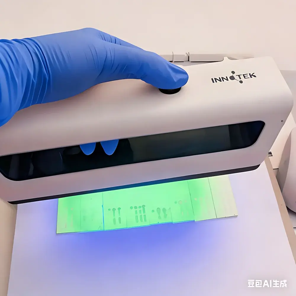
The MIBIscope Multi-Ion Beam Imaging Mass Spectrometer is a high-performance secondary ion mass spectrometry (SIMS) platform engineered for spatially resolved, multiplexed proteomic imaging of formalin-fixed paraffin-embedded (FFPE) and frozen tissue sections. Leveraging multi-ion beam imaging (MIBI) technology, the system employs a focused, high-current oxygen primary ion beam (30 keV) to sputter molecular fragments and metal-tagged antibodies from tissue surfaces. Emitted secondary ions are analyzed via time-of-flight mass spectrometry with high mass resolution (11,000–16,000) and extended mass range (>12,000 u), enabling simultaneous detection of over 40 metal-conjugated antibodies per acquisition. Unlike conventional immunofluorescence or single-beam SIMS systems, MIBIscope delivers quantitative, subcellular-resolution protein distribution maps without spectral overlap—by exploiting the isotopic purity and narrow mass peaks of rare-earth lanthanide tags (e.g., 113In, 166Er). This architecture supports robust, reproducible spatial proteomics in translational research, tumor microenvironment characterization, and biomarker validation workflows aligned with CLIA- and GLP-informed laboratory practices.
Key Features
- Multi-ion beam raster scanning with real-time ion yield normalization and beam current stabilization for consistent signal intensity across large-area acquisitions (up to 800 × 800 µm2 ROI)
- Time-of-flight mass analyzer with reflectron configuration, delivering mass resolution ≥11,000 (FWHM) at m/z 150–200 and linear dynamic range exceeding 104 (5-log)
- Integrated optical and scanning electron detector (SED) imaging for precise region-of-interest (ROI) selection and co-registration with histological context
- Automated antibody tag calibration using certified reference standards traceable to NIST SRM 2917 (lanthanide oxide nanoparticles)
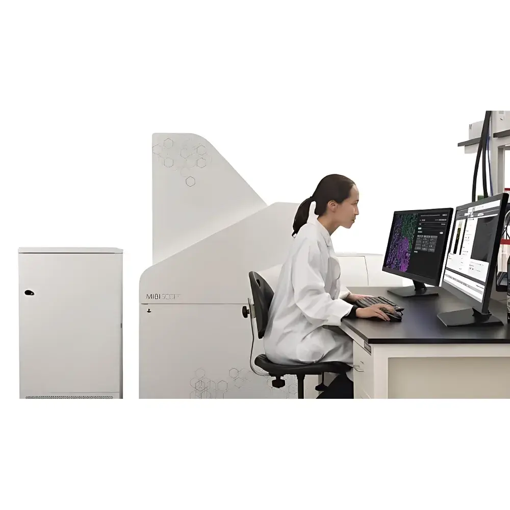
- Turnkey operation via intuitive GUI; no dedicated mass spectrometry expertise required—designed for integration into routine pathology lab environments
- Low maintenance architecture: cryo-pumped ultra-high vacuum chamber (<1×10⁻⁹ mbar), solid-state ion source, and field-replaceable detector modules
Sample Compatibility & Compliance
The MIBIscope accepts standard glass-mounted tissue sections (4–10 µm thickness), including FFPE, OCT-embedded frozen, and touch-prep samples. Compatible with standard metal-tagged antibody panels (Fluidigm/MAXpar® or custom conjugates), it supports validated staining protocols compliant with CAP/CLIA pre-analytical guidelines. Data acquisition and processing adhere to ISO/IEC 17025:2017 requirements for analytical competence, and raw data files (TIFF-based hyperstack format) retain full metadata—including instrument parameters, calibration timestamps, and operator logs—for audit readiness under FDA 21 CFR Part 11 and GLP/GMP frameworks.
Software & Data Management
Acquisition and analysis are managed through MIBI Analyst v4.x, a validated software suite supporting automated ROI definition, spectral deconvolution, pixel-wise quantification, and spatial clustering (PhenoGraph, HotSpot Analysis). All processed datasets export to open formats (OME-TIFF, HDF5) compatible with downstream tools including R/Bioconductor (Cytofkit, imcRtools), Python (scikit-image, napari), and commercial platforms (Visiopharm, HALO). Audit trails record every parameter change, user action, and calibration event; electronic signatures and role-based access control ensure compliance with regulatory data integrity standards.
Applications
- High-parameter tumor immune profiling: simultaneous mapping of PD-1/PD-L1, CD3/CD4/CD8, FOXP3, Granzyme B, and macrophage markers within spatial neighborhoods
- Developmental biology: 3D reconstruction of protein gradients in embryonic tissue sections using serial section alignment
- Therapeutic response monitoring: longitudinal assessment of target engagement and pathway modulation in xenograft biopsies
- Biomarker discovery: unsupervised segmentation of rare cell phenotypes (e.g., stem-like or exhausted T cells) correlated with clinical outcomes
- Neuropathology: multiplexed visualization of amyloid-beta isoforms, tau phospho-epitopes, and glial activation markers in Alzheimer’s disease brain sections
FAQ
What sample preparation protocols are validated for MIBIscope?
Standard MAXpar® antibody staining on antigen-retrieved FFPE sections or methanol-fixed frozen sections is fully supported; protocol SOPs are available under NDA.
Can MIBIscope perform depth profiling or 3D imaging?
Yes—sequential ablation with in situ surface reconstruction enables layer-by-layer 3D volume acquisition up to ~20 µm depth with isotropic voxel resolution (350 nm × 350 nm × 500 nm).
Is raw data export compatible with third-party image analysis pipelines?
All acquisitions generate OME-TIFF hyperstacks with embedded metadata; Python and R APIs are provided for batch processing and integration with existing bioinformatics infrastructure.
What level of service and support is included post-installation?
Comprehensive 3-year platinum support package includes remote diagnostics, annual performance verification (per ASTM E1941), on-site application training, and priority access to software updates and method development consulting.