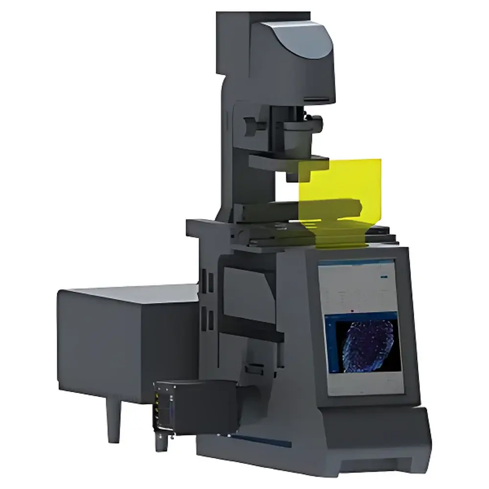
Micro-Field HM-SR-01 STORM Super-Resolution Fluorescence Microscope
| Brand | Micro-Field / Guangzhou Micro-field Optical Technology Co., Ltd. |
|---|---|
| Origin | Guangdong, China |
| Manufacturer Type | Authorized Distributor |
| Product Category | Domestic |
| Model | HM-SR-01 |
| Imaging Modality | dSTORM (direct Stochastic Optical Reconstruction Microscopy) & TIRF (Total Internal Reflection Fluorescence) |
| Objectives | Plan-Apochromat 40×/0.95 (WD: 0.25–0.16 mm) |
| Apochromat 100×/1.45 Oil (WD | 0.13 mm) |
| Excitation Lasers | 405 nm (60 mW), 432 nm (80 mW), 488 nm (20 mW), 561 nm (80 mW), 638 nm (60 mW) |
| Focus Stability | Active autofocus with ≤50 nm axial drift correction |
| Z-Stage | Closed-loop nanopositioning stage (150 µm travel, 0.8 nm resolution) |
| XY-Stage | Motorized stage (110 × 75 mm travel, ±1 µm repeatability) |
| Camera Interface | High-sensitivity scientific CMOS or EMCCD compatible |
| Dimensions | 840 × 345 × 750 mm |
Overview
The Micro-Field HM-SR-01 STORM Super-Resolution Fluorescence Microscope is a rigorously engineered platform designed for quantitative nanoscale fluorescence imaging in fixed and live-cell biological specimens. It implements two complementary high-resolution modalities: direct Stochastic Optical Reconstruction Microscopy (dSTORM) and Total Internal Reflection Fluorescence (TIRF) microscopy. Unlike widefield or confocal systems limited by the diffraction barrier (~250 nm lateral resolution), the HM-SR-01 achieves isotropic spatial resolution down to ~20 nm through single-molecule localization—leveraging photoswitchable fluorophores, precise laser excitation control, and statistical reconstruction algorithms. Its dual-mode architecture enables users to select between evanescent-field–confined surface imaging (TIRF) for membrane-proximal dynamics and stochastic single-molecule blinking (dSTORM) for dense, sub-diffraction structural mapping—both fully integrated within a single optical train. The system was developed in alignment with national instrumentation R&D initiatives, emphasizing robustness, reproducibility, and operational accessibility without compromising on metrological fidelity.
Key Features
- Dual-modal optical architecture supporting both dSTORM and TIRF imaging via manual or motorized light-path switching—enabling seamless transition between surface-restricted live-cell dynamics and ultra-high-resolution structural reconstruction.
- Optimized optical train incorporating Plan-Apochromat 40×/0.95 and Apochromat 100×/1.45 oil-immersion objectives, each corrected for chromatic and spherical aberration across visible and near-UV wavelengths—critical for multi-color dSTORM registration and minimal PSF distortion.
- Multi-wavelength TIRF illumination system with five independently controllable solid-state lasers (405 nm, 432 nm, 488 nm, 561 nm, 638 nm), coupled via dichroic combiner and calibrated TIRF angle control—ensuring consistent evanescent field depth (≤100 nm) and minimal background.
- Closed-loop nanopositioning Z-stage with 0.8 nm encoder resolution and 150 µm travel range—providing sub-nanometer axial stability during prolonged acquisitions required for single-molecule localization.
- Real-time active autofocus system utilizing infrared reflection-based focus lock, maintaining axial precision ≤50 nm over multi-hour sessions—essential for mitigating thermal drift and mechanical perturbation in long-term super-resolution time-lapse experiments.
- Integrated central touchscreen interface for coordinated control of illumination, stage, filter wheels, and acquisition parameters—reducing software layer complexity and minimizing user-induced configuration errors.
Sample Compatibility & Compliance
The HM-SR-01 supports standard glass-bottom dishes (e.g., MatTek, ibidi), coverslips (No. 1.5H), and custom sample chambers compatible with aqueous or oil-immersion mounting. It accommodates live-cell imaging under environmental control (CO₂, temperature, humidity) when paired with external incubation enclosures. All optical components meet ISO 10110 surface quality standards; objective lenses comply with ISO 8578 for numerical aperture tolerance and working distance specification. The system’s hardware and firmware architecture support audit-trail–capable operation in GLP-compliant environments; data acquisition metadata (timestamp, laser power, exposure, stage position, objective ID) is embedded in TIFF/OME-TIFF headers per OME-NGFF conventions. While not pre-certified for FDA 21 CFR Part 11, the platform permits integration with validated third-party LIMS or ELN systems for regulated workflows.
Software & Data Management
The HM-SR-01 ships with Micro-Field SR-Studio—a modular acquisition and analysis suite built on Qt/C++ with Python API extension support. Acquisition modules include real-time TIRF alignment assistant, dSTORM blinking optimization wizard, and multi-channel registration calibration tool. Localization analysis employs maximum-likelihood estimation (MLE) and Bayesian drift correction algorithms compliant with the ASU Super-Resolution Microscopy Standards Consortium guidelines. Processed data exports to standardized formats: CSV (localization lists), HDF5 (multi-dimensional stacks), and OME-Zarr (for cloud-native analysis pipelines). Raw image streams are stored in lossless TIFF with embedded EXIF and custom metadata tags—including laser wavelength, power, exposure time, and objective magnification—for traceability and FAIR data principles adherence.
Applications
- Nanoscale mapping of synaptic protein organization (e.g., PSD-95, Bassoon) in primary neuronal cultures using multi-color dSTORM.
- Quantitative analysis of clathrin-coated pit dynamics at the plasma membrane via TIRF time-lapse with frame rates up to 100 fps.
- Sub-diffraction characterization of nuclear pore complex distribution and stoichiometry in fixed mammalian nuclei.
- Co-localization analysis of receptor dimerization (e.g., EGFR, PDGFR) with <10 nm precision in cancer cell lines.
- Correlative imaging workflows integrating HM-SR-01 with atomic force microscopy (AFM) or fluorescence resonance energy transfer (FRET) setups—enabled by shared XYZ coordinate referencing and synchronization TTL triggers.
FAQ
What sample preparation protocols are recommended for dSTORM imaging on the HM-SR-01?
Standard immunolabeling with Alexa Fluor 647, CF680, or ATTO 655 conjugates is advised. Use of the included Micro-Field Super-Resolution Buffer Kit ensures optimal photoswitching kinetics, low background, and pH stability over >30 min acquisition windows.
Is the system compatible with third-party cameras?
Yes—the HM-SR-01 provides standard USB 3.2 Gen 2 and Camera Link interfaces, supporting scientific CMOS (e.g., Hamamatsu ORCA-Fusion BT) and EMCCD (e.g., Andor iXon Ultra) sensors with trigger synchronization and ROI readout control.
Can TIRF and dSTORM modes be used simultaneously?
No—optical path selection is mutually exclusive via physical beam-splitter positioning; however, rapid mode switching (<5 s) allows sequential acquisition within the same session without realignment.
Does the autofocus system require fluorescent reference beads?
No—it operates via infrared reflection from the coverslip-sample interface, eliminating need for fiducial markers or bead embedding that may perturb biological integrity.
What maintenance is required for long-term stability?
Annual calibration of laser power output, objective alignment, and Z-stage encoder linearity is recommended. No routine optical realignment is needed due to monolithic optomechanical baseplate design and passive thermal stabilization.


