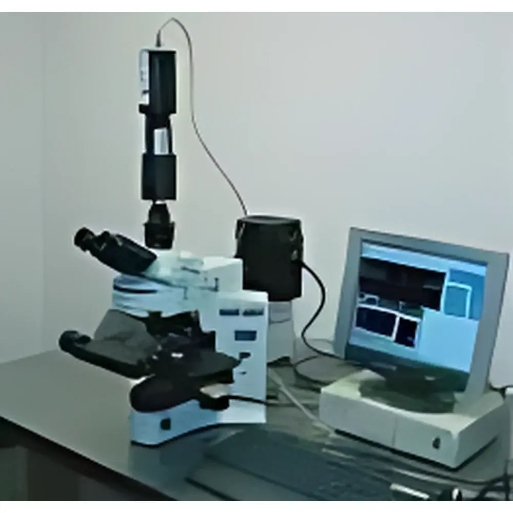
MicroHSImager Hyperspectral Microscopy System
| Origin | Finland |
|---|---|
| Manufacturer Type | Authorized Distributor |
| Origin Category | Imported Instrument |
| Model | MicroHSImager |
| Pricing | Available Upon Request |
Overview
The MicroHSImager Hyperspectral Microscopy System is a precision-engineered integrated platform that combines high-resolution optical microscopy with push-broom or snapshot-based hyperspectral imaging technology. Designed specifically for life science and biomedical research laboratories, this system captures spatially resolved spectral data across contiguous narrowband wavelength channels—typically spanning the visible to near-infrared (VNIR) range (400–1000 nm)—enabling label-free, non-destructive biochemical characterization at the cellular and subcellular level. Unlike conventional RGB or fluorescence microscopy, the MicroHSImager acquires full reflectance, transmittance, or absorbance spectra (e.g., 128–256 bands at 2–5 nm spectral resolution) for every pixel in the field of view, generating a three-dimensional data cube (x, y, λ). This architecture supports quantitative spectral unmixing, spectral library matching, and multivariate statistical analysis—critical for identifying endogenous chromophores (e.g., hemoglobin, melanin, collagen), detecting pathological tissue margins, or mapping drug distribution in ex vivo histological sections.
Key Features
- Integrated microscope-compatible design with standardized C-mount or infinity-corrected optical interface for seamless coupling to upright or inverted research-grade microscopes.
- VNIR hyperspectral sensor with configurable spectral range (default: 450–950 nm) and adjustable spectral sampling interval (2.5 nm typical), optimized for biological contrast without external illumination artifacts.
- High spatial fidelity maintained via diffraction-limited optics and pixel-aligned spectral calibration; supports magnifications from 4× to 63× objective lenses with real-time spectral registration.
- Motorized XYZ stage with sub-micron repeatability for automated tile-scanning of large-area tissue sections or multi-well plates.
- Thermoelectrically cooled scientific CMOS detector ensuring low dark current and high signal-to-noise ratio (SNR > 60 dB) under extended acquisition times.
- Rugged aluminum-alloy chassis with EMI-shielded electronics, compliant with IEC 61000-6-3 and IEC 61000-6-4 emission standards for shared laboratory environments.
Sample Compatibility & Compliance
The MicroHSImager accommodates standard glass slides (1 mm thickness), coverslips (0.13–0.17 mm), frozen or FFPE tissue sections, cell monolayers on transparent substrates, and 3D organoid cultures in imaging-compatible chambers. It operates without exogenous dyes or tags, preserving native spectral signatures—a key advantage for longitudinal studies and regulatory-compliant preclinical evaluation. The system meets ISO/IEC 17025 requirements for measurement traceability when used with NIST-traceable reflectance standards. Data acquisition workflows support GLP-compliant documentation: metadata embedding (sample ID, operator, timestamp, objective lens, exposure time), audit trail logging, and export in HDF5 or ENVI-compatible formats for third-party validation.
Software & Data Management
Controlled via SpectraView™ Pro software (v4.2+), the platform provides real-time preview, spectral calibration (using built-in tungsten-halogen and diffuse reflectance references), and batch processing pipelines. Advanced analytical modules include PCA-based anomaly detection, MCR-ALS spectral unmixing, and supervised classification (SVM, Random Forest) trained on user-defined ground-truth regions. All processed results—including false-color spectral maps, concentration heatmaps, and spectral libraries—are exportable as TIFF, CSV, or MATLAB .mat files. Software adheres to FDA 21 CFR Part 11 requirements: electronic signatures, role-based access control, and immutable audit logs for raw data modification events.
Applications
- Label-free histopathology: Discrimination of tumor infiltration zones, necrotic cores, and stromal interfaces in unstained tissue sections.
- Cellular metabolism monitoring: Quantification of redox ratios (NADH/FAD) via autofluorescence spectral decomposition in live-cell imaging.
- Pharmaceutical development: Spatial mapping of active pharmaceutical ingredient (API) crystallinity and polymorphic form distribution within tablet cross-sections.
- Neuroscience research: Visualization of myelin lipid composition gradients in white matter tracts using vibrational overtone absorption features.
- Plant phenotyping: Detection of early-stage biotic stress responses through chlorophyll-a/b ratio shifts and carotenoid degradation signatures.
FAQ
What spectral range does the MicroHSImager cover by default?
The standard configuration operates in the visible to near-infrared (VNIR) range from 450 nm to 950 nm, with optional extensions into SWIR (950–1700 nm) available upon request.
Is the system compatible with existing microscope brands?
Yes—it integrates with major platforms including Zeiss Axio, Leica DMi8, Nikon Eclipse Ti2, and Olympus BX series via standardized mechanical and optical interfaces.
Can spectral data be exported for use in MATLAB or Python environments?
Absolutely. Raw and processed data are exportable in HDF5, ENVI BIL, and CSV formats, with documented APIs for direct integration into SciPy, scikit-learn, or TensorFlow workflows.
Does the system require external calibration before each session?
No—integrated reference sources enable one-click radiometric and spectral calibration; routine verification is recommended daily using NIST-traceable standards.
How is data integrity ensured during long-term deployment in regulated labs?
Through built-in 21 CFR Part 11 compliance features: electronic signature enforcement, encrypted audit trails, and version-controlled analysis scripts with hash-verified outputs.


")