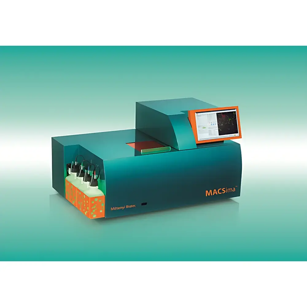
Miltenyi Biotec MACSima™ Automated Spatial Profiling Imaging System
| Brand | Miltenyi Biotec |
|---|---|
| Origin | Germany |
| Manufacturer | Yes |
| Import Status | Imported |
| Model | MACSima |
| Pricing | Upon Request |
Overview
The Miltenyi Biotec MACSima™ Automated Spatial Profiling Imaging System is an integrated, fully automated platform engineered for high-plex, spatially resolved protein profiling in fixed biological specimens. It implements the proprietary MACSima Imaging Cyclic Staining (MICS) technology—a fluorescence-based, iterative immunostaining and signal erasure methodology rooted in sequential epitope labeling, high-fidelity image acquisition, and non-destructive fluorophore inactivation. Unlike conventional multiplex immunofluorescence or mass cytometry approaches that face spectral overlap or tissue degradation constraints, MICS enables quantitative detection of >100 distinct protein targets on a single tissue section or cell monolayer—preserving spatial architecture and antigen integrity across cycles. The system operates on a robust optical architecture featuring precision motorized stage control, high-sensitivity sCMOS imaging, and hardware-synchronized illumination, delivering subcellular resolution with consistent z-stack registration across all staining rounds. Designed for translational research and preclinical biomarker discovery, MACSima supports hypothesis-driven and exploratory spatial biology workflows without requiring specialized computational infrastructure at the point of acquisition.
Key Features
- Fully automated end-to-end workflow: experimental design → cyclic staining → image acquisition → signal erasure → data export — executed without manual intervention
- Dual-mode fluorophore erasure: enzymatic cleavage and photochemical inactivation pathways validated for >200 recombinant REA-conjugated antibodies
- MACSwell™ sample carrier system: three standardized formats (tissue slide, adherent cell chamber, suspension cell well) with defined reaction volumes and thermal stabilization for reproducible antigen accessibility
- Integrated real-time QC metrics: cycle-by-cycle signal-to-noise ratio monitoring, focus drift correction, and autofluorescence subtraction embedded in acquisition firmware
- REAscreen™ pre-validated antibody panels: off-the-shelf, lot-controlled fluorescent antibody cocktails optimized for MICS compatibility and minimal cross-reactivity
- Hardware-level synchronization: LED excitation control, filter wheel timing, and stage positioning coordinated at microsecond resolution to ensure pixel-perfect registration across cycles
Sample Compatibility & Compliance
MACSima accepts formalin-fixed paraffin-embedded (FFPE) tissue sections (4–10 µm), frozen sections, cytospins, and cultured adherent or suspension cells—provided samples are appropriately fixed (e.g., 4% PFA or methanol-acetone) and antigen-retrieved per validated protocols. All reagents—including REA-conjugated antibodies, erasure buffers, and mounting media—are CE-IVD marked and compliant with ISO 13485 quality management systems. The platform supports GLP-aligned documentation: full audit trail logging (user actions, timestamps, instrument parameters), electronic signatures per FDA 21 CFR Part 11 requirements, and raw image metadata retention (TIFF/OME-TIFF) compliant with MIAME and MINSEQE reporting standards. Validation packages include inter-laboratory reproducibility data per ISO/IEC 17025 guidelines for spatial protein quantification.
Software & Data Management
Data acquisition and primary processing are managed by MACS® iQ Control software, which enforces protocol versioning, cycle parameter locking, and automatic backup to network-attached storage (NAS) or cloud endpoints via encrypted SFTP. MACS® iQ View provides modular analysis modules: spatial clustering (Phenograph, Leiden), neighborhood analysis (Cell Neighborhood Mapping), expression gradient modeling, and co-expression correlation heatmaps—all operating natively on registered 3D image stacks without down-sampling. Export options include annotated OME-Zarr containers, CSV matrices with spatial coordinates (x, y, z, marker intensity), and interactive HTML reports compatible with downstream tools (QuPath, HALO, Visium-compatible formats). All software components undergo annual penetration testing and maintain SOC 2 Type II certification for data handling integrity.
Applications
- Tumor microenvironment mapping: simultaneous quantification of immune checkpoint proteins (PD-1/PD-L1/CTLA-4), stromal markers (α-SMA, FAP), and tumor lineage antigens within intact tissue architecture
- Neurodegenerative disease phenotyping: spatial colocalization of phosphorylated tau isoforms, Aβ plaques, microglial activation states, and synaptic density markers in human post-mortem brain sections
- Developmental biology: time-resolved protein gradient analysis across embryonic tissue sections using serial section alignment and morphometric registration
- Immunotherapy response prediction: longitudinal spatial profiling of T-cell infiltration patterns, exhaustion markers, and antigen-presenting cell maturation states in paired pre-/post-treatment biopsies
- Single-cell spatial transcriptomics integration: anchor-based registration of MACSima protein maps with Visium or Xenium RNA data using shared structural landmarks (nuclei, vasculature)
FAQ
What is the maximum number of markers achievable per sample?
Up to 150+ protein targets have been validated in peer-reviewed studies using REAscreen™ panels and dual-erasure optimization; practical throughput depends on antibody validation status and tissue antigen density.
Does MACSima require specialized training for operation?
Operators require foundational knowledge in immunohistochemistry and digital pathology; however, the system’s guided workflow interface and context-sensitive help reduce hands-on training to under 8 hours for certified lab personnel.
Can MACSima data be integrated with third-party analysis platforms?
Yes—raw OME-TIFF and processed CSV/JSON exports are interoperable with QuPath, HALO, CellProfiler, and Python-based spatial analysis libraries (squidpy, scanpy, spatialmuon).
Is MACSima compatible with multiplexed RNA detection methods?
While MACSima is protein-centric, its coordinate-mapped output serves as a spatial scaffold for integrating in situ hybridization or sequencing-derived RNA data via landmark-based registration algorithms.
How is data integrity ensured during long-duration cyclic runs?
The system performs hourly internal calibration checks (focus stability, LED intensity drift, stage repeatability) and logs all deviations; any deviation exceeding ±0.5 µm positional error triggers automatic pause and alert notification.


