Neoscan N80 Desktop High-Resolution Micro-CT System for Agricultural and Forestry Research
| Brand | Neoscan |
|---|---|
| Origin | Belgium |
| Detector Type | Flat-Panel Detector |
| Scan Mode | Sample Rotation Only (RO) |
| Spatial Resolution | 2 µm |
| X-ray Energy | 110 kV |
| Field of View | 100 mm × 163 mm |
| Maximum Sample Weight | 20 kg |
| System Dimensions | 1200 mm × 640 mm × 520 mm |
| Precision | Micron-Level |
Overview
The Neoscan N80 Desktop High-Resolution Micro-CT System is an industrial-grade, non-destructive 3D X-ray imaging platform engineered for quantitative structural and density analysis of biological and porous materials in agricultural and forestry research. Based on cone-beam computed tomography (CBCT) principles, the system employs a microfocus X-ray source and a high-dynamic-range flat-panel detector to acquire hundreds of projection images as the sample rotates on a precision air-bearing stage. Reconstruction algorithms—such as filtered back-projection (FBP) and iterative regularization methods—generate isotropic volumetric datasets with true voxel sizes down to 2 µm, enabling sub-cellular visualization of plant tissues, root architecture, seed morphology, and soil pore networks without physical sectioning or staining. Unlike synchrotron-based systems, the N80 delivers laboratory-accessible, benchtop-operable performance with stable tube output and thermal management optimized for long-duration scans (up to 24 h), making it suitable for longitudinal studies of root growth dynamics or soil hydration processes under controlled environmental conditions.
Key Features
- Micron-level spatial resolution (≤2 µm) achieved via geometric magnification and high-resolution flat-panel detector (≥2048 × 2048 pixels, 12-bit dynamic range)
- 110 kV microfocus X-ray source with adjustable current (10–200 µA) and focal spot size <5 µm, supporting dual-energy contrast enhancement for material differentiation
- Sample rotation-only (RO) scanning geometry ensures mechanical stability and reproducibility; no moving source/detector components reduce alignment drift and maintenance requirements
- Integrated high-precision rotary stage with angular accuracy ≤0.01° and load capacity up to 20 kg, accommodating intact soil cores, potted plants, or harvested stems
- Benchtop footprint (1200 × 640 × 520 mm) with lead-shielded enclosure compliant with IEC 61000-6-3 and EU Directive 2013/59/Euratom for radiation safety
- Real-time preview mode with live reconstruction latency <2 s per slice, enabling rapid parameter optimization prior to full acquisition
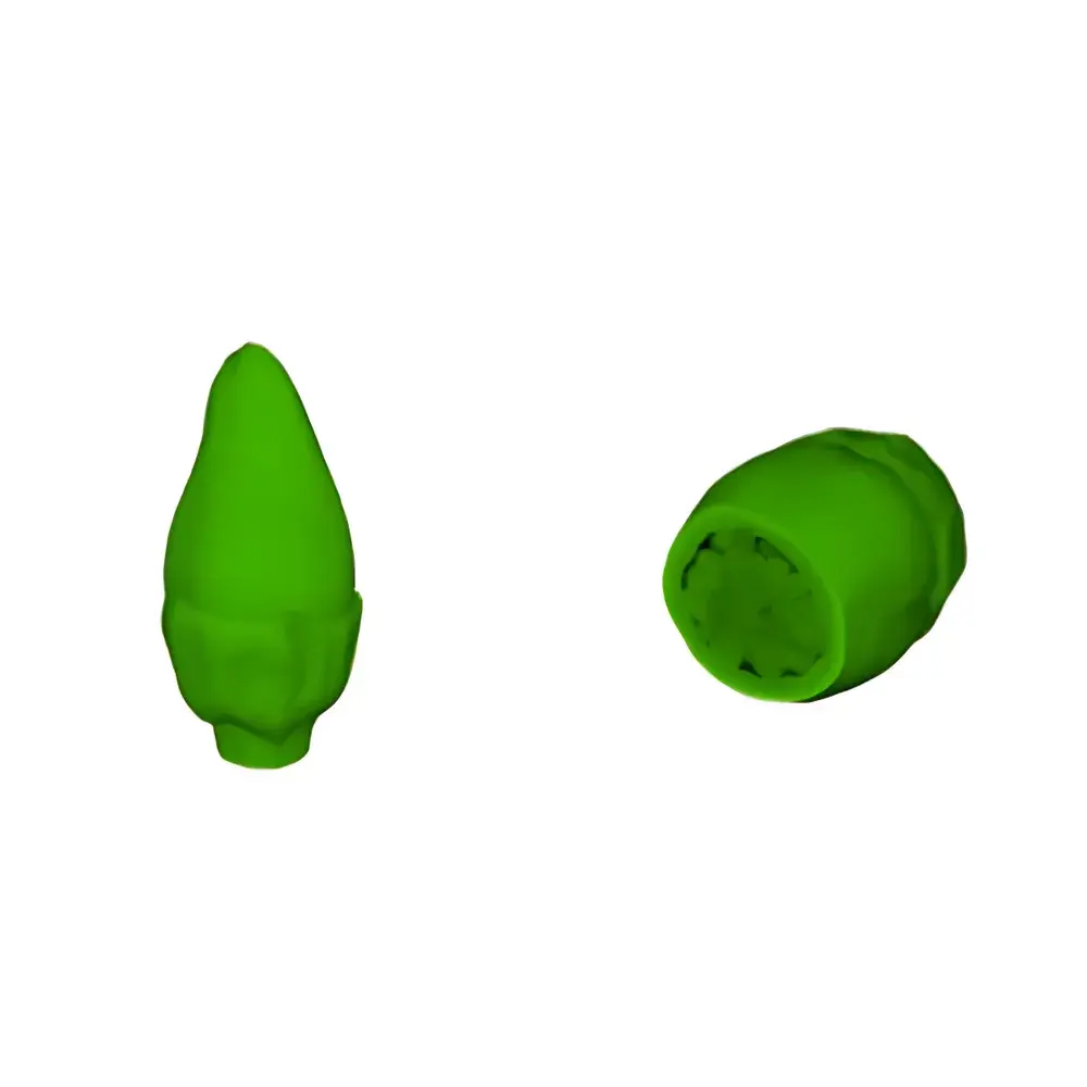
Sample Compatibility & Compliance
The N80 supports a broad spectrum of agronomic specimens—including intact seed units (e.g., rice, tomato, maize), excised root systems embedded in soil or hydrogel matrices, woody stem cross-sections, leaf petioles, fungal hyphae colonizing substrates, and undisturbed soil monoliths (up to Ø100 mm × H163 mm). Its low-dose acquisition protocols minimize radiation-induced artifacts in living or hydrated samples, preserving viability for post-scan phenotyping. Data outputs conform to DICOM 3.0 and TIFF stack formats, facilitating interoperability with third-party segmentation tools (e.g., Avizo, Dragonfly, BoneJ). The system meets ISO 15739:2013 for noise measurement in digital radiography and ASTM E1441-22 for computed tomography standard practice. For regulated environments, optional audit trail logging and user access controls align with GLP/GMP documentation requirements and support FDA 21 CFR Part 11 compliance when deployed with validated software modules.
Software & Data Management
The N80 operates with Neoscan’s proprietary CT Suite v4.x, a modular application suite comprising acquisition control, GPU-accelerated reconstruction (CPU + CUDA hybrid), and semi-automated segmentation workflows tailored for botanical structures. Built-in morphometric engines quantify porosity distribution, specific surface area, tortuosity, connectivity density, and skeletonized root length metrics directly from binary volumes. Batch processing pipelines enable unattended multi-sample runs with metadata tagging (e.g., genotype, treatment group, timepoint). Export options include STL for 3D printing, VTK for finite element mesh generation, and CSV tables compatible with R, Python (scikit-image, SimpleITK), and MATLAB. All raw projections and reconstructed volumes are stored with embedded EXIF-like headers containing exposure parameters, geometric calibration coefficients, and timestamped operator logs—ensuring traceability across experimental replicates.
Applications
- Plant phenomics: Quantitative 3D characterization of vascular bundle patterning in cereal stems, grain chalkiness distribution in milled rice, and embryo viability assessment in dormant seeds
- Root-soil interface analysis: In situ visualization of root penetration pathways, rhizosphere pore topology changes during drought/rehydration cycles, and mycorrhizal colonization mapping
- Soil structure quantification: Segmentation of aggregate hierarchy, pore network modeling for hydraulic conductivity estimation (per Kozeny-Carman correlation), and organic matter spatial distribution analysis
- Crop breeding support: High-throughput screening of internal traits (e.g., endosperm void fraction, pericarp thickness, hilum integrity) as non-destructive selection markers
- Pathology & entomology: Detection of larval galleries within wood or fruit tissue, fungal hyphal infiltration routes in leaves, and gall formation dynamics without dissection
FAQ
What is the minimum achievable voxel size for biological soft-tissue samples?
The system achieves effective isotropic voxel sizes down to 2 µm under optimal magnification and signal-to-noise conditions; actual resolution depends on sample attenuation, scan duration, and reconstruction kernel selection.
Can the N80 perform time-lapse (4D) imaging of growing roots?
Yes—integrated environmental chamber compatibility (optional) enables temperature- and humidity-controlled sequential scanning over days; motion correction algorithms compensate for minor stage drift between acquisitions.
Is phase-contrast or edge-enhancement capability available?
While not a synchrotron source, the N80 leverages propagation-based phase effects at high geometric magnifications (>10×) and applies Paganin-type filter reconstruction to enhance soft-tissue boundaries in low-contrast specimens like young root tips.
How does the system handle beam hardening artifacts in heterogeneous samples like soil-root composites?
CT Suite includes dual-energy pre-correction, polynomial-based hardening compensation, and iterative reconstruction with total variation regularization to suppress cupping and streaking artifacts.
Are hardware upgrades (e.g., higher-power source, larger detector) supported post-purchase?
Neoscan designs the N80 with modular subsystem interfaces; detector and source modules are field-replaceable under service contract, with firmware and reconstruction software updated to maintain compatibility.


